
Fig. 5.
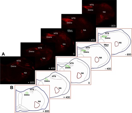
Nucleus ambiguus. A: montages of confocal projections of right brain stem sections showing NA motoneurons (−800 to + 800 μm relative to obex) that were retrogradely labeled by tetramethylrhodamine dextran (TMR-D) injection into the right nodose ganglion in a RA rat. B: brain stems were traced, scaled, and superimposed, using a Neurolucida tracing system to define the boundaries of the NA at each level, as enclosed in the red contours (n = 6). Note: vagal afferent terminals in the nucleus of the solitary tract (NTS) and vagal motoneurons in the DmnX were also labeled by TMR-D.