
Fig. 6.
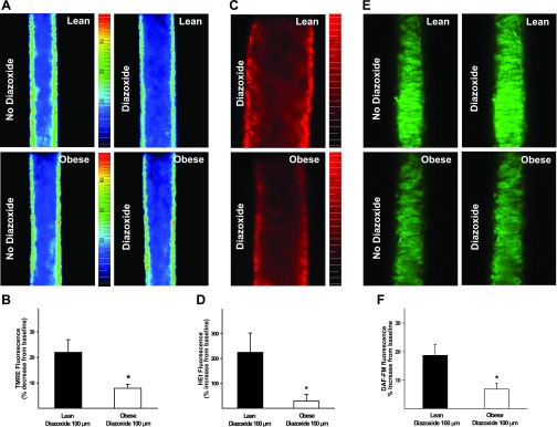
Membrane depolarization, reactive oxygen species (ROS) generation, and nitric oxide (NO) production in isolated pressurized cerebral arteries of ZO and ZL rats before and after diazoxide (100 μmol/l) administration.A: fluorescence images of the mitochondrial membrane potential-sensitive dye tetramethylrhodamine ethyl ester (TMRE) in ZL (top) and ZO (bottom) arteries in the absence and presence of diazoxide. B: TMRE fluorescence response to diazoxide. Reduction of diazoxide-induced decrease in TMRE fluorescence in ZO compared with ZL arteries indicates reduced mitochondrial depolarization. C: fluorescence images of the ROS sensitive dye hydroethidine (HEt) in ZL (top) and ZO (bottom) arteries following diazoxide administration. D: HEt fluorescence response to diazoxide compared with vehicle. E: fluorescence images of the NO-sensitive dye 4-amino-5-methylamino-2′,7′-difluorofluorescein diacetate (DAF-FM) in ZL (top) and ZO (bottom) arteries in the absence and presence of diazoxide. F: DAF-FM fluorescence response to diazoxide compared with vehicle. Reduction of diazoxide-induced increase in HEt and DAF-FM fluorescence in ZO compared with ZL arteries indicates decreased generation of ROS and NO, respectively. *Significantly different from lean (P < 0.05).