Fig. 1.
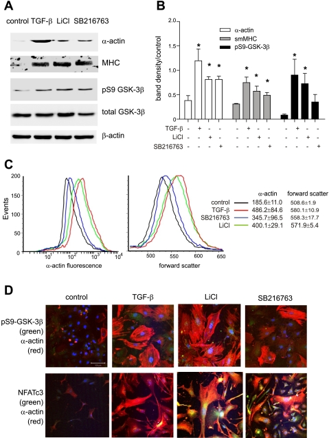
Glycogen synthase kinase-3β (GSK-3β) inhibition increases cell size and contractile protein synthesis in cultured murine airway smooth muscle (ASM) cells. A: mouse ASM cells from control animals were serum-deprived and treated with 10 ng/ml transforming growth factor (TGF)-β or the GSK-3β inhibitors 10 mM LiCl or 50 nM SB-216763. After 3 days of treatment, samples were processed for Western blots using antibodies to α-actin, myosin heavy chain (MHC), phospho-Ser9 GSK-3β (pS9 GSK-3β), total GSK-3β, and β-actin. B: group mean data for immunoblots as quantified by densitometry. Data are represented as fold increase over control. Data from 3 individual experiments are shown (*P < 0.05). C: mouse ASM cells were untreated (black line) or treated with 10 ng/ml TGF-β (red line), 50 nM SB-216763 (blue line), or 10 mM LiCl (green line). Cells were trypsinized, immunostained with anti-α-actin-FITC, and processed for flow cytometry. TGF-β, SB-216763, and LiCl each significantly increased cell size (as measured by forward scatter) and α-actin immunostaining (*P < 0.05, 1-way ANOVA; n = 3). D: fluorescence confocal microscopy. Cells were treated with vehicle, TGF-β, LiCl, or SB-216763 immunostained with rabbit anti-pGSK (green channel, top 3 frames) or anti-NFATc3 (green channel, bottom 3 frames) and anti-α-actin-Cy3 (red channel). Nuclei were counterstained with Hoechst 33342 (blue channel). Merged images are shown. In the cytoplasm, colocalization of pGSK or NFATc3 with α-actin is orange to yellow. Localization of pGSK or NFATc3 in the nucleus is pale blue (without α-actin) or white (with α-actin). Both TGF-β and GSK-3β inhibitors increased cell size, pGSK content, nuclear localization of NFAT, and α-actin expression. The white bar is 100 μm, and all images are to the same scale. sm, Smooth muscle.

