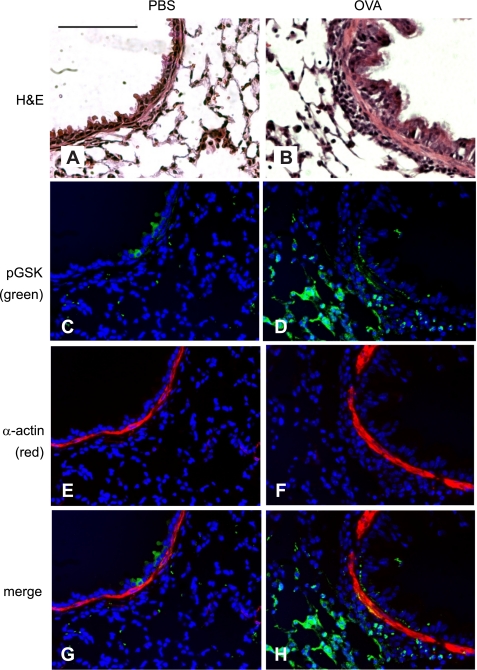
Fig. 4.
OVA treatment increases phospho-Ser9 GSK-3β (pGSK) immunostaining in the airway epithelium and ASM. A and B: H&E-stained lung sections from PBS- and OVA-treated mice. C–F: lung sections were immunostained with anti-phospho-GSK-3β (green, C and D) or anti-α-actin Cy3 conjugate (red, E and F). Nuclei were stained with Hoechst 33342 (blue, C–H). Images were merged (G and H) to show colocalization of phospho-GSK-3β and α-actin. Colocalization varies in intensity from orange (relatively low pGSK) to yellow (higher pGSK). OVA-treated mice (B, D, F, and H) showed increased airway inflammation (B), phospho-GSK-3β (D), α-actin (F), and colocalization of phospho-GSK-3β and α-actin (H). Magnification is ×320, black bars represent 50 μm, and all panels are to the same scale.

