Fig. 6.
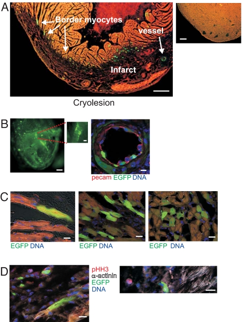
Reexpression of c-kit-EGFP after injury in the adult heart. (A) (Left) Stefanini-fixed heart 7 days postsurgery. Note the large number of EGFP+ cells at the border zone and within the infarcted (darker) area. (Right) Rare fluorescent cells in sham heart. (B) EGFP cells within infarct. (Left) Fluorescence wide-field image of adult c-kitBAC-EGFP heart 14 days postcryoablation. Note fluorescent cells lining vessel. (Right) c-kit-EGFP+ endothelial cells lining an artery 7 days postinjury are identified by PECAM1 staining; surrounding smooth muscle cells also express EGFP. (C) EGFP+ cardiomyocytes at border zone. (Left) Fluorescent and nonfluorescent striated cardiomyocytes. (Center and Right) Clusters of EGFP+ myocytes at border zone 7 days postinfarct. (D) Lack of pHH3 in EGFP+ cardiac myocytes. (Left) EGFP+ and EGFP− myocytes at border zone. (Right) Single pHH3+ nonmyocyte within infarct. (Scale bars: A Left, 360 μm; A Right, 280 μm; B Left, 250 μm; B Inset, 100 μm; B Right, 10 μm; C, 22 μm; D Left, 14 μm; D Right, 16 μm.)

