Fig. 3.
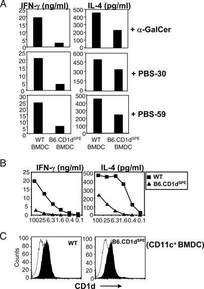
Impaired type I NKT cell activation by exogenous glycolipid antigens presented by B6.CD1dSPE BMDC. (A) Purified type I NKT cells were stimulated with WT or B6.CD1dSPE BMDC pulsed with the antigen indicated. The amount of IFN-γ and IL-4 was quantitated by ELISA. Bar graphs depict mean values from duplicate wells. The data shown are representative of 2 independent experiments. (B) Line graph depicts IFN-γ and IL-4 production by Vα14-transgenic TCRα knockout splenocytes in response to WT and B6.CD1dSPE BMDC pulsed with titrating concentrations of α-GalCer. Data shown are representative of 3 independent experiments. (C) Histograms depict CD1d expression on WT and B6.CD1dSPE BMDC used in the assay. BMDC were stained with mAb to CD11c and a control mAb (open) or mAb to CD1d (filled) and analyzed by flow cytometry.

