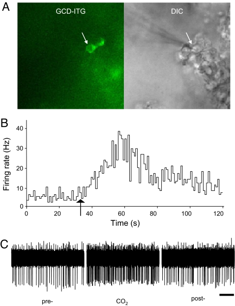
Fig. 1.
CO2 activates GC-D+ neurons. (A) Targeted recording of GC-D+ neurons. GC-D+ cells were identified by green fluorescence in both somata and knobs in an olfactory epithelial preparation from GCD-ITG mice (Left). An electrode was guided toward a GC-D+ cell (arrows) by using DIC microscopy (Right) for cell-attached extracellular recording. (B) The mean firing rate of a GC-D+ cell was dramatically enhanced after CO2 application in the perfusion solution (arrow). (C) Sample physiological traces showing the firing of the neurons shown in B before (pre), during (CO2), and after CO2 application (post). (Scale bar, 2 s.)

