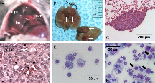
Figure 2.
Malignant pleural effusions and pleural tumors in C57B/6 mice after intrapleural LLC propagation. (A) Transdiaphragmatic view of a malignant pleural effusion (mpe), a supradiaphragmatic tumor mass (tm), and the heart (h). (B) Lung and chest wall explants showing multiple tumor foci on the parietal and visceral pleura (arrows). (C) Section through a small visceral pleural Lewis lung cancer implantation stained with hematoxylin and eosin shows tumor growth on the pleural surface (Å = 100). (D) New vessel formation within a pleural LLC tumor (Å = 400, arrowheads = new vessels formed within tumor tissue). (E) Cytological preparation (cytospin) from pleural fluid obtained from an untreated mouse stained with modified Wright's-Giemsa stain (Å = 400). (F) Representative photomicrograph of a cytospin from a malignant pleural effusion stained with modified Wright's-Giemsa stain (Å = 400). LLC cells with large nuclei and visible nucleoli (arrows) can be observed among with a variety of host inflammatory cells.

