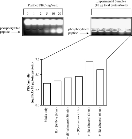
Figure 8.
PKC activity in NHBE cells is increased by (R)-albuterol. Cells were stimulated with 10 ng/ml each of IL-1β and IFN-γ for 6 h. (R)-albuterol (10−5 M) was added to cytokine-stimulated cultures at the indicated time points, at which time cells were lysed and immediately assayed for PKC activity. (R)-albuterol appeared to augment PKC activity maximally at 3 h exposure, followed by a diminution of the effect. Band intensity reflects phosphorylation activity by PKC, which was quantified by imaging software (UVP, Ltd., Upland, CA). Values were extrapolated from a linear standard curve based on a series dilution of purified PKC supplied with the assay kit (gel on left). Data are representative of three individual experiments.

