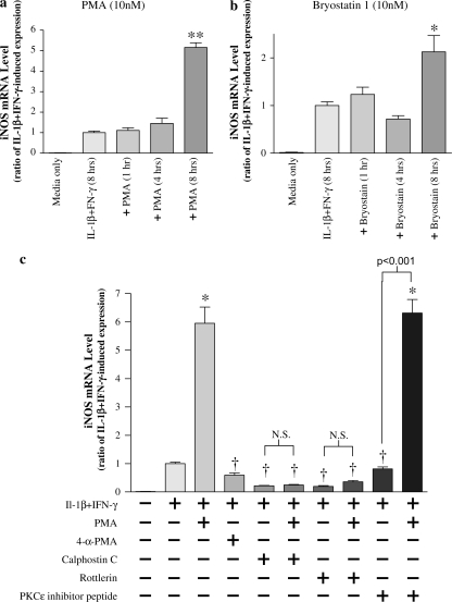
Figure 9.
iNOS gene expression is reversibly enhanced by PKC activators. (a) NHBE cells were stimulated with 10 ng/ml each of IL-1β and IFN-γ for 8 h. The general PKC activator phorbol 12-myristate 13-acetate (PMA, 10 nM), or the specific PKCδ/ε activator bryostatin 1 (10 nM), were added for 1, 4, and 8 h, at which times iNOS message levels also were measured. Both PMA and bryostatin 1 augmented iNOS message at 8 h. Data are presented as mean ± SEM (n = 6). * Significantly more than cytokine-induced iNOS expression (P < 0.01, ** = P < 0.001). (b) PMA (10 nM) was added to NHBE cells for 8 h in the presence of either control medium, calphostin C (500 nM), rottlerin (3 μM), or PKCε translocation inhibitor peptide (300 μM), and iNOS message was assessed. In selected wells, the PMA negative control, 4α-PMA (10 nM), was added in place of PMA. The increase in iNOS message in response to PMA was attenuated by either calphostin C or rottlerin, suggesting that PKCδ was involved. Neither the PKCε translocation inhibitor nor 4α-PMA affected iNOS expression. PKC activation alone with bryostatin 1 (10 nM) in the absence of IL-1β + IFN-γ did not affect iNOS expression (data not shown). Data are presented as mean ± SEM (n = 4–8). * Significantly greater than cytokine-induced iNOS expression (P < 0.001); † significantly lower than cytokine + PMA–induced iNOS expression (P < 0.001). N.S., no significant difference (P > 0.05).

