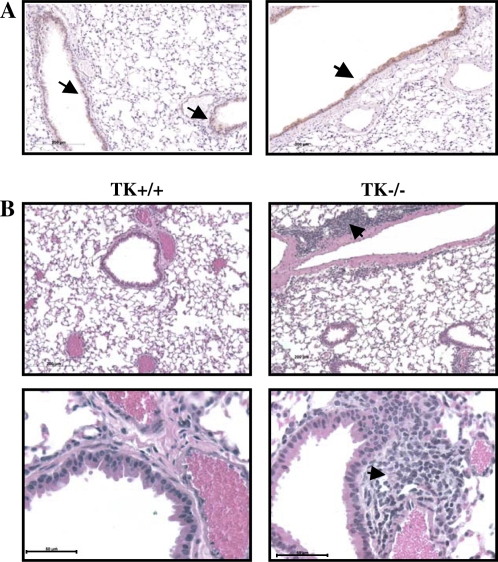
Figure 7.
Histology of lung sections from untreated mice. (A) Sections from the lungs of two different Mst1r TK+/+ mice were stained with an anti-Mst1r antibody and counterstained with hematoxylin. Arrows indicate the presence of Mst1r around the airway epithelium of the lungs. (B) Sections from the lungs of Mst1r TK+/+ and Mst1r TK−/− mice were stained with hematoxylin and eosin Y. Clusters of cells are observed in the lungs of TK−/−, but not wild-type mice. These clusters are observed in all of the TK−/− mice examined.

