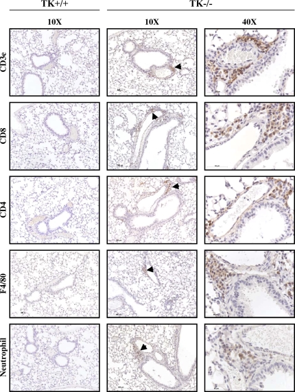
Figure 8.
Leukocytes in lung tissue of Mst1r TK−/− mice. Tissue sections from the lungs of Mst1r TK+/+ and TK−/− mice were stained for various inflammatory cell markers. No staining was observed in the TK+/+ lungs. Positive staining in the TK−/− lungs indicates the presence of T cells (CD3e, CD4, and CD8), natural killer cells (CD8), macrophages (F4/80), and neutrophils in the cell clusters.

