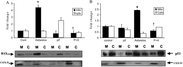
Figure 5.
Asbestos-induced A549 cell mitochondrial Bax (A) and p53 (B) translocation is reduced by pifithrin. A549 were exposed to asbestos (25 μg/cm2) in the presence or absence of pifithrin (pif) for 24 h. Mitochondrial and cytosolic protein were obtained and assessed for Bax (A) and p53 (B) protein by Western analysis. The differences observed in the levels of Bax and p53 from three experiments are shown in a densitometric analysis of mitochondrial (M; solid bars) and cytosolic (C; open bars) proteins. The levels of cytochrome oxidase IV (COXIV) were used to confirm the presence of mitochondrial protein and comparable loading. Data are expressed as mean ± SEM of control value. *P < 0.05 versus control; †P < 0.05 versus asbestos. P+A: pifithrin and asbestos.

