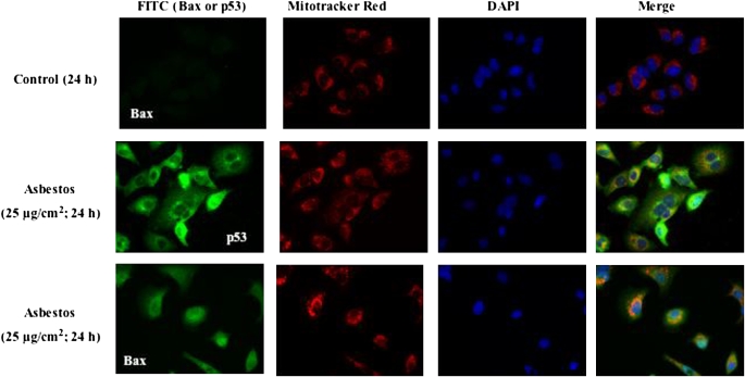
Figure 6.
Unlike controls, asbestos induces mitochondrial colocalization of p53 and Bax. A549 cells were exposed for 24 h to control medium (first row) or asbestos (25 μg/cm2; second and third rows), and mitochondrial accumulation of p53 (second row) and Bax (third row) were assessed by confocal immunofluorescence. After exposure, the cells were collected and triple-stained for mitochondria (Mito-tracker Red), nuclei (DAPI), and pro-apoptotic molecule (p53 or Bax; FITC-PoAb). Asbestos-induced mitochondrial localization of p53 and Bax are evident as the yellow-orange punctate cytoplasmic/perinuclear staining in the merged samples (fourth column). There is also evidence of p53 and PUMA nuclear staining in some cells.

