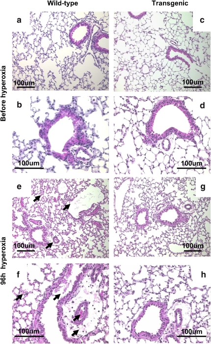
Figure 3.
Lung morphology with hyperoxia. Lungs were fixed, sectioned, and stained with hematoxylin and eosin. Lungs of Prdx6 Tg (c, d) and age-matched wild-type mice (a, b) showed similar histologic appearance before oxygen exposure. With 96 h exposure to hyperoxia, wild-type mice (e, f) compared with Prdx6 Tg mice (g, h) showed greater degree of tissue injury (arrows) with perivascular edema, hemorrhage, cellular necrosis, and cellular infiltration. Photomicrographs are representative of n = 3 in each group. The scale bar is 100 μm.

