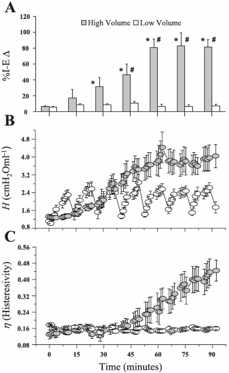
Figure 6.
A) Percent change in open alveoli between the end of inspiration and the end of expiration (%I-EΔ) observed by in vivo microscopy in rats receiving either high volume (injurious) or low volume (protective) mechanical ventilation. %I-EΔ in the high-volume group increases progressively with time indicating an increase in the fraction of alveoli that collapse with each expiration, while %I-EΔ does not change in the low-volume group indicating no accrual of lung tissue damage. B) Corresponding measurements of the parameter H (Eq. 2), an index of lung stiffness. C) Corresponding measurements of the ratio of G to H, termed hysteresivity. In the low-volume rats, even thought H increases transiently until reversed by periodic deep lung inflations, hysteresivity does not change with time, indicating that gradual but reversible derecruitment of alveoli was responsible for the changes in H. By contrast, hysteresivity increased progressively and irreversibly in the high-volume rats, indicating that their lung tissues were becoming increasingly damaged with time (reproduced with permission from (Allen et al., 2005).

