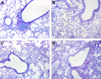
Figure 6.
Lung histology. Sections of the lungs were prepared 48 h after infection with 5 × 10⁶ cfu of P. aeruginosa. There was more infiltration by inflammatory cells in the lungs of SP-A−/− (B), SP-D−/− (C), and SP-AD−/− (D) mice compared with WT mice (A). These sections are representative of three different animals per genotype.

