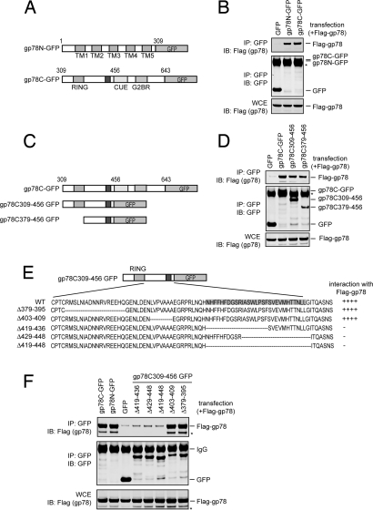
Fig. 1.
Oligomerization of gp78. (A) Schematic representation of the gp78 variants tested in B. (B) Both the cytosolic domain and the transmembrane segments of gp78 can interact with full-length gp78. Detergent extracts of 293T cells transfected with the indicated plasmids were subjected to immunoprecipitation (IP) followed by immunoblotting (IB) with the indicated antibodies. Note that the expressed gp78 proteins comigrate with IgG (*). WCE, whole cell extract. (C–F) Mapping the region in the gp78 cytosolic domain that is necessary for its self-association. (C) Schematic representation of the gp78 variants tested in D. (D) As in B, except that plasmids expressing the indicated gp78 variants were analyzed. (E) Schematic representation of the gp78 variants used in F. (F) As in B, except that plasmids expressing the indicated gp78 variants were analyzed. * indicates a gp78 degradation product.

