Fig. 5.
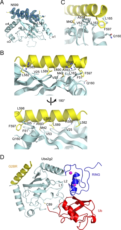
The structure of Ube2g2 bound to the gp78 G2BR domain. (A) Overall structure showing the Ube2g2–G2BR complex. Contoured at 3.5 σ around G2BR is a 2Fo − Fc σA-weighted annealed omit map omitting G2BR. (B) The contacts between G2BR and Ube2g2. (C) A close-up view on the most critical contacts around A593 and F597 of G2BR. (D) The geometry of G2BR binding compared with RING binding and the active site. RING domain (blue) of the c-Cbl–UbcH7 complex (Protein Data Bank ID code 1FBV), and Ub (red) of the Mms2-Ubc13∼Ub covalent complex (Protein Data Bank ID code 2GMI) are docked onto the Ube2g2–G2BR complex based on E2 structural alignments. Overlay matrices were determined by DALI.

