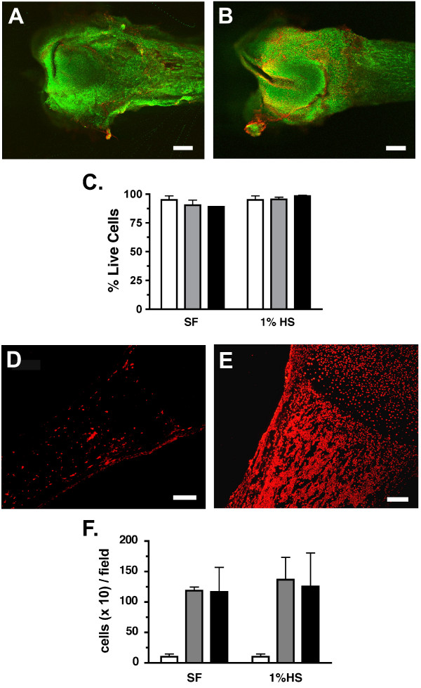
Figure 2.
Viability of femur tissue in culture. A & B. Photomicrograph of femurs freshly dissected (A) or cultured in 1% horse serum (HS) for 2 days (B). Images are merged z-stacked slices through ~250 μM of the outer surface of the bone. Scale bars equal 0.38 mm (A & B). C. % live (calcein green) cells in femurs cultured in serum free (SF) or 1% horse serum (1% HS) for 0 days (fresh; white bar), 2 days (gray) and 4 days (black). Data represents an average of 3 femurs per treatment and time point. D & E. Photomicrographs of sections taken from freshly dissected femurs assayed for the presence of apoptotic cells (red) within the diaphysis and the marrow cavity (D). Adjacent sections (E) were treated with DNase I prior to TMR staining as described. Scale bars equal 150 μm. F. Average number of apoptotic cells per field counted within the diaphysis is shown for fresh BN sections (white bar) and sections from BN cultured for 2 days (gray) or 4 days (black) in SF or 1% HS media.

