Figure 7.
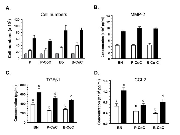
Cocultures of breast carcinoma cells and mouse femurs show changes in MCP-1 secretions. The human breast adenocarcinoma parental cell line, MDA-MB-231, and the boneseeking subclone known as MDA-MB-231 BO, were cultured alone and with mouse femurs. Viability of these cells in culture, as assessed by cell numbers, and analysis of secreted factors was conducted as described. Note that no detectable expression of these factors was observed in single cultures of parental or bone subclones. A: Cell numbers (× 103) are shown for the parental line (P), the parental-femur coculture (P-CoC), the bone subclone (Bo) and the Bo-femur coculture (B-CoC) at the initial plating (gray bar) and day 1 (white) and day 2 (black) of culture. B-D: Concentration of MMP2 (B), TGFβ1 (C), and MCP-1 (D) is shown for the femur alone (BN) and the cocultures at day 1 (white) and day 2 (black) of culture. In C & D, significant differences between concentration of the secreted factors observed in BN cultures (a or c) compared to cocultures with parental or bone-specific MDA cells is indicated by a different letter above the Co-C bars, with significance at P < 0.01 (b) or P < 0.001 (d). Across three independent experiments, media was sampled from 7–8 wells for each time point, culture condition and media.

