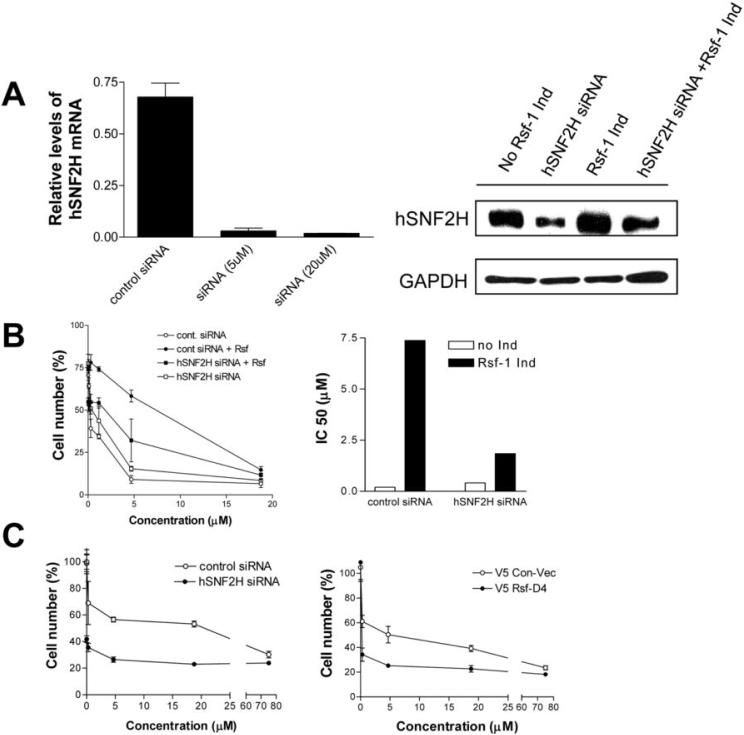
Figure 4. The role of hSNF2H in paclitaxel sensitivity.
A. Quantitative RT-PCR shows mRNA expression levels of hSNF2H are greatly reduced by hSNF2H siRNA in OVCAR3 cells with high level expression of endogenous hSNF2H (left panel). Similarly, Western blot analysis shows reduction of hSNF2H expression by hSNF2H siRNA 24 hours after siRNA transfection in the Rsf-1 inducible SKOV3 cells (right panel). siRNA against luciferase was used as the control. B. Rsf-1 inducible cells transfected with control or hSNF2H siRNAs were treated with various concentrations of paclitaxel under Rsf-1 turned-on or -off condition. Relative cell number in different treatment group was determined and presented as percentage of the control group at day 3 (left panel). IC50 of paclitaxel in Rsf-1 inducible SKOV3 cells transfected with control or hSNF2H siRNAs were determined under Rsf-1 turned-on or -off condition (right panel). hSNF2H siRNAs significantly decreases the IC50 of paclitaxel in Rsf-1 expressing SKOV3 cells. C. Effect of hSNF2H siRNA and Rsf-D4 expression on sensitivity of OVCAR3 cells to paclitaxel. In contrast to SKOV3 cells, OVCAR3 cells constitutively express Rsf-1 due to a high level of 11q13.5 amplification. Both hSNF2H siRNA (left panel) and Rsf-D4 (right panel) significantly sensitize OVCAR3 cells to paclitaxel treatment. The cell number at each concentration points was normalized to hSNF2H siRNA and Rsf-D4 treated cells without paclitaxel treatment.

