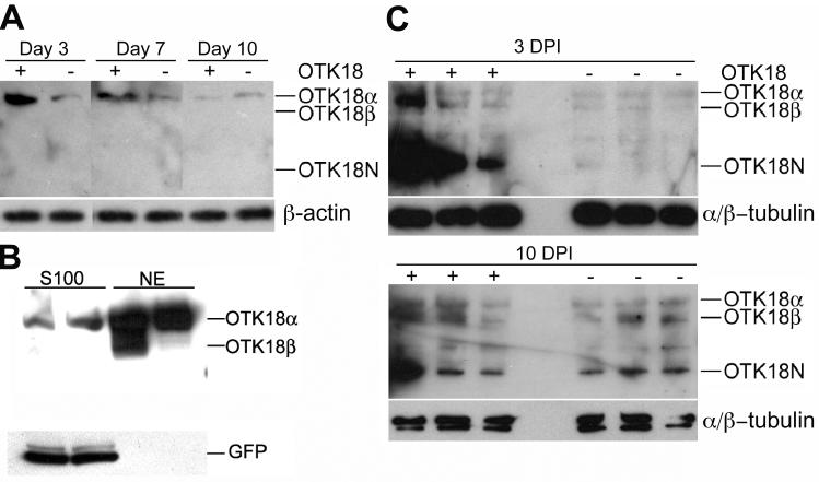
Figure 1. Processing of OTK18 upon HIV-1 infection of MDM.
A, Immunoblotting of OTK18 (upper panel) and β-actin (lower panel) 3, 7, and 10 days after Ad-OTK18/GFP (+) or Ad-GFP (-) adenovirus infection using anti-OTK18 mAb specific to OTK18 1-180 amino acids. Only OTK18α (75 kD), but neither OTK18β (65 kD) nor OTK18N (35 kD), was detected. B, Immunoblotting of cytoplasmic (S100) and nuclear extract (NE) of OTK18-expressing MDM after subcellular fractionation. 40 μg of each fraction was loaded onto 4-15% SDS-PAGE and subjected to immunoblotting using anti-OTK18 (upper panel) or anti-GFP (lower panel) monoclonals. OTK18N was undetected in these fractions. C, Immunoblotting of OTK18 (upper panel) and α/β-tubulin (lower panel) at 3 and 10 days post HIV infection (DPI). Thirty million MDM were infected with adenovirus (Ad-OTK18/GFP (+) or Ad-GFP adenovirus (-)), followed by infection with HIV-1ADA (one day later). OTK18α (75 kD), OTK18β (65 kD) and OTK18N (35 kD) were detected in both Ad-OTK18/GFP and Ad-GFP-infected MDM at Day 10.

