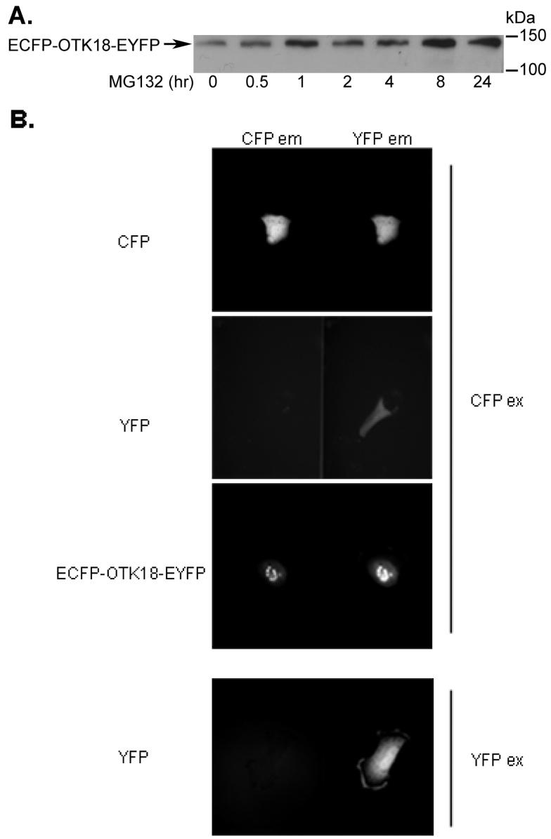
Figure 6. Expression and FRET analysis of ECFP-OTK18-EYFP.
A, The cells were transfected with ECFP-OTK18-EYFP and treated with 10 mM MG132 for the times indicated. Total cell extract was subjected to immunoblotting using anti-OTK18 mAb. B, The cells were transfected with ECFP, EYFP, or ECFP-OTK18-EYFP 24 hours prior to imaging, and excited with CFP filter (CFPex for ECFP, EYFP, or ECFP-OTK18-EYFP) or YFP filter (EYFP only), and images were taken by CFP filter (CFPem, left panel) and YFP filter (YFPem, right panel) using Dual-View imaging system. Original magnification: 400X.

