
Fig. 3.
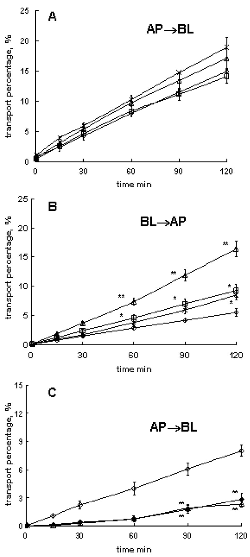
Effects of P85 on 3H-Phe transport in different directions across BBMEC monolayers: a AP to BL transport of 3H-Phe with/without P85 at the AP side (isolation 1), b BL to AP transport of 3H-Phe with/without P85 at BL side (isolation 2), c AP to BL transport of 3H-Phe with/without P85 or 2-mM unlabeled Phe at BL side (isolation 3). The treatment groups included: a, b assay buffer controls (open diamonds), 0.01 wt.% P85 (crosses), 0.1 wt.% P85 (open triangles), and 1 wt.% P85 (open squares) and c assay buffer (open diamond), 0.1% P85 (open triangle), and 2-mM unlabeled Phe in assay buffer (filled diamond). BBMEC monolayers were grown on 12-well transwell inserts. Statistical comparisons were made pair-wise between (1) each P85 and (2) assay buffer groups: p<0.05 (*) and p<0.01 (**), n=3