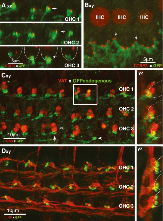
FIG. 2.
GABAB1 receptors are expressed by terminals of type I and type II ganglion cells on IHCs or OHCs, respectively. Anti-GFP (green) is used to enhance the endogenous signal from the GABAB1-GFP reporter, except in the inset of C, where the endogenous GFP signal is shown. Images are 2-D projections from confocal z-stacks of cochlear whole mounts. The projection view is indicated in each panel: xy surface view; yz cross-section view; xz view from tunnel of Corti. In several panels, the approximate positions of OHCs are indicated by dashed lines. A, B Proximity of GABAB1-positive terminals on OHCs (A) or IHCs (B) to synaptic ribbons immunostained with CtBP2 (red). Axz projections through four OHCs from each of the three rows. Filled arrows indicate terminal clusters closely associated with synaptic ribbons. Bxy projection of three IHCs from 20 serial images spanning 5 μm in the z dimension. Arrows point to two synaptic ribbons. C GABAB1 containing terminals on OHCs (filled arrow) are complementary to OC terminals (unfilled arrow) expressing GAD (red). D Neurofilament antibody (NF-200; red) reveals spiraling type II axons terminating in GABAB1-positive terminals under three rows of OHCs.

