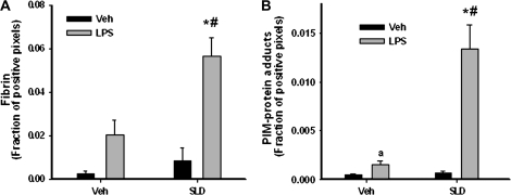
FIG. 6.
Evaluation of fibrin deposition and hypoxia in liver. Rats were treated as described in Figure 2. (A) Animals were killed at 4 h, and livers were processed for immunohistochemical determination of fibrin deposition. (B) Rats received an additional treatment with PIM hydrochloride (120 mg/kg) 2 h after the second administration of SLD. They were then killed at 4 h, and livers were processed for immunohistochemical determination of hypoxia. In both panels the fraction of positive pixels averaged from 10 randomly chosen microscope fields (100×) was determined for each animal. *Significantly different from Veh/LPS group. #Significantly different from SLD/Veh group. aSignificantly different from Veh/Veh group. p < 0.05, n = 4–6.

