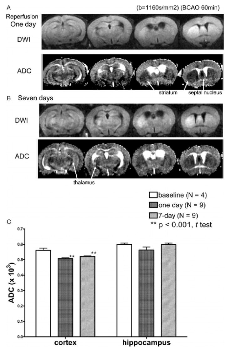
Figure 3.

Comparison of diffusion-weighted imaging (DWI) and apparent diffusion coefficient (ADC) between bilateral carotid artery occlusion (BCAO) and sham-operated animals at two time points: DWIs and ADC were obtained from animals 1 (A) and 7 (B) days after treatment with BCAO of 60 minutes. The ADCs (mean ± SD) in sham-operated (baseline) animals were 0.57 ± 0.01 × 10⁻³ and 0.61 ± 0.01 × 10⁻³ for the cortex and the hippocampus, respectively. Regions with edema can be identified by comparing the ADC of the 1- and 7-day time points with baseline ADC (C). Edema, identified by visual inspection of DWI hyperintensity and reduced ADC, occurred in the striatum and septal nucleus in all animals but in the thalamus and hypothalamus of only four of nine animals.
