
Figure 7.
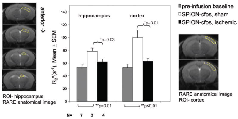
Profiles of superparamagnetic iron oxide nanoparticle (SPION) retention in R2* maps of three groups of animals. Probe retention in the ischemia-treated group is significantly lower than in the sham-operated group (t-test). No significant difference in retention was observed between baseline animals without SPION-cfos infusion and cerebral ischemia–treated animals with SPION-cfos infusion. The number of animals is given below the bar graph. The outlines of the hippocampus and cortex were referenced from the anatomic images and superimposed on individual R2* maps as regions of interest (ROI); R2* values within two ROIs of each animal were extracted from individual R2* maps as ROI-hippocampus and ROI-cortex for statistical analysis. Mean R2* and standard error (SEM) are shown. RARE = rapid acquisition with relaxation enhancement.