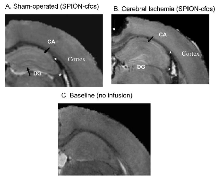
Figure 8.

Less cerebral superparamagnetic iron oxide nanoparticle (SPION) retention in animals with stroke cerebral ischemia. Three-dimensional T2*-weighted images of postmortem brains collected 3 days after intracerebroventricular infusion of SPION-cfos and treatment with either sham operation (A) or bilateral carotid occlusion (B) for 60 minutes 1 week before. The hippocampus of the contralateral hemisphere is shown from one representative mouse in each group. Short arrows show the dentate gyrus (DG) and pyramidal cell layer (CA) neuronal formation in the hippocampus. C shows an animal brain without infusion. The signal void in the upper-left corner of B (thin arrow) was caused by missing tissue during brain sample handling. The number of animals is listed in Figure 7.
