
Figure 1.
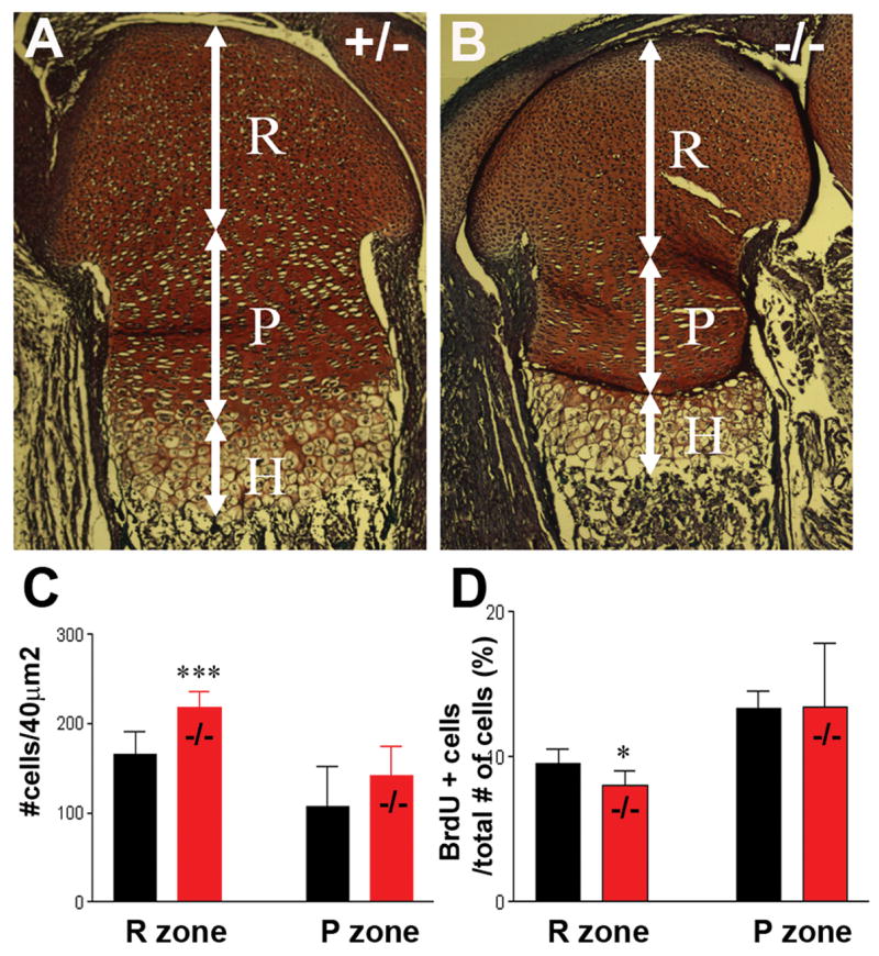
Cell density is increased in the reserve zone of HIP/RPL29 null epiphyses of distal embryonic femurs. Although the epiphysis/growth plates of d18.5 embryos (B) were shorter (white arrows) compared with controls (A), no overall alteration of cartilage organization was observed. However, the layer of proliferating (P) chondrocytes was shortened, and a significant increase in cellular density was seen in the reserve zone (R) of null embryos (C). BrdU-labeling revealed a small but significant decrease in chondroprogenitor proliferation in the R zone of developing nulls bones compared to controls (D). *** and * denote statistical significance differences (p < 0.001 and p < 0.05, respectively).