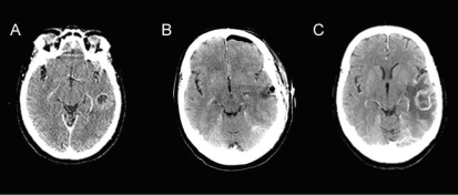
FIGURE 1.
Post-contrast computed tomography (A) before resection demonstrates a part-solid, part-cystic, ring-enhancing lesion in the left temporal lobe; (B) immediately following resection demonstrates a resection cavity containing air and fluid without an enhancing abnormality (left frontal postsurgical pneumocephalus); (C) 2 months post-resection demonstrates expansion of ring enhancement within the left temporal lobe, associated with increased perilesion edema and mass effect.

