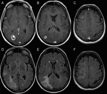
FIGURE 5.
Magnetic resonance imaging slices showing glioblastoma multiforme (case 2). (A–C) Axial T1 post-gadolinium images and (D–F) corresponding axial flair (fluid-attenuated inversion recovery) slices at the same levels. Ring-enhancing masses can be seen in (A) the right temporal lobe, (A) right occipital lobe, (B) right inferior parietal lobe, (C) right frontal lobe, and (C) right superior parietal lobe. Under flair, hyperintensity can be seen around each lesion (D–F). Additional flair hyperintensity is seen intervening between the right frontal lobe and the right superior parietal lobe lesions (F).

