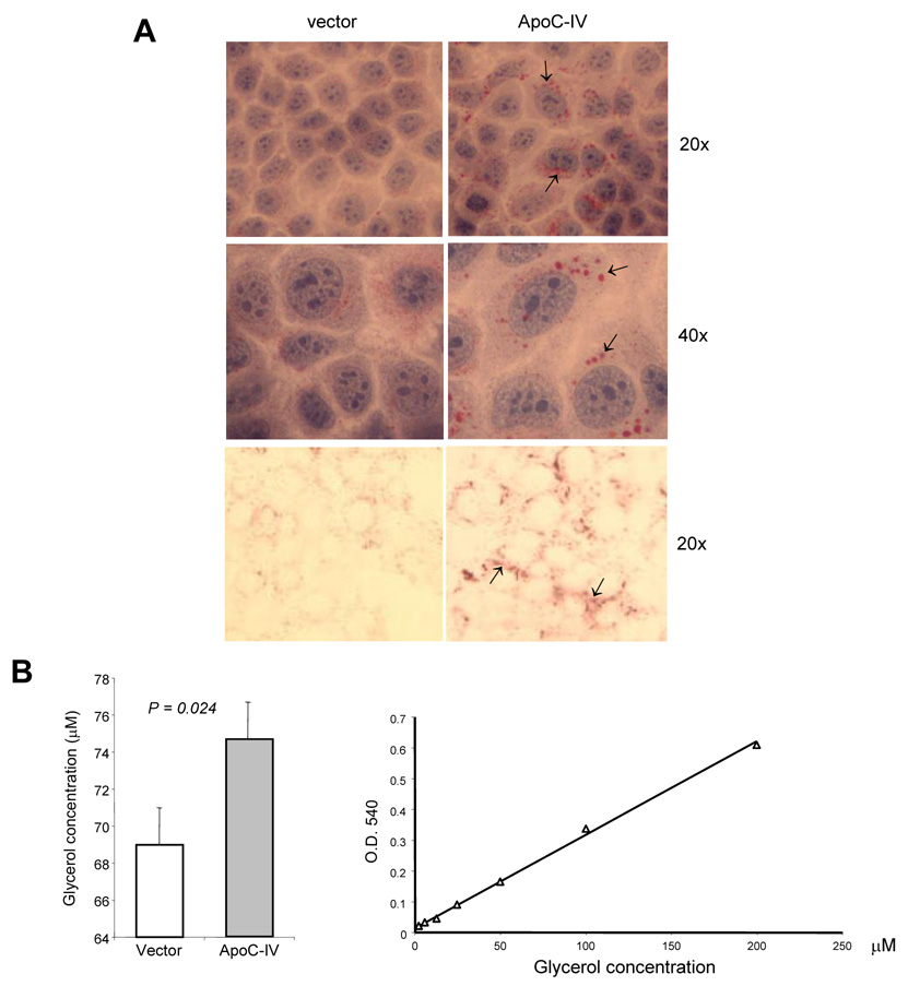
FIG. 7. Triglyceride accumulation in transfected Huh-7 cells.
(A) Oil Red O staining of cells transfected with the ApoC-IV cDNA or empty vector. Upper and middle panels: cells cultured in FBS-free medium with additional H&E staining. Lower panel: cells cultured in delipidated medium. Arrows indicate lipid accumulation. (B) Enzymatic assay of triglyceride in transfected cells (left panel) and standard curve (right panel).

