Figure 6.
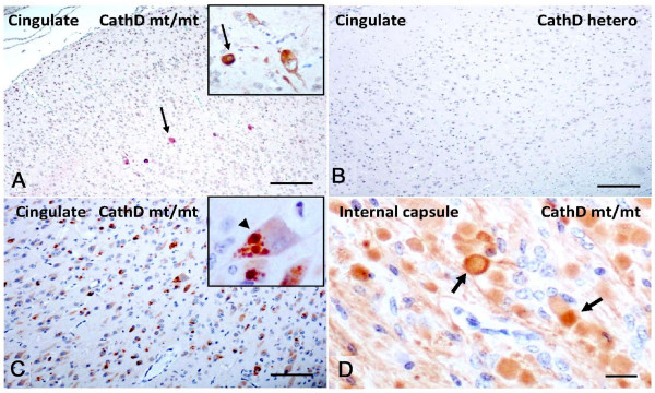
Sheep expressing a homozygous mutant of Cathepsin D exhibit accumulation of phosphorylated α-synuclein in cingulate cortex. (A) Select cortical neurons (arrow) of cingulate gyrus in homozygous mutants (CathD mt/mt) show cytoplasmic immunostaining with Ser129-phosphorylated aSyn Ab. (B) Absence of Ser129-phosphorylated aSyn-reactivity in the cingulate gyrus of age-matched, heterozygous CTSD (CathD hetero) sheep brain. (C) Anti-ubiquitin antibody staining reveals extensive, often multiple, intra-cellular inclusion formation in neurons (arowhead) of affected sheep throughout the cortex including cingulate cortex. (D) Immunostaining for aSyn with polyclonal Ab, hSA-2, reveals abundant aSyn deposits and axonal swelling (arrows) in the internal capsule of homozygous mutant sheep but not wild-type (not shown) or heterozygous sheep (not shown). Scale bars, 100 μm in A, B, C, and 15 μm in D.

