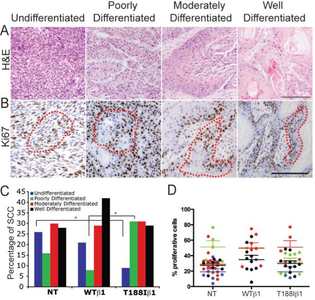
Figure 6. Different types of malignant tumour.

Sections of malignant tumours labelled with H&E (A) or anti-Ki67 (brown) with haematoxylin counterstain (B). Red dotted lines in (B) demarcate proliferative regions quantitated in (D). Scale bars: 100 μm. (C, D) Quantitation of proportion of each tumour type (C) and % Ki67 positive cells within the proliferative regions of each tumour type (D). NT: non-transgenic WTβ1: K14WTβ1 transgenic; T188Iβ1: K14T188Iβ1 transgenic. * indicates p < 0.05 (Pearson’s chi-square test).
