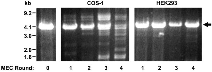
Figure 4.
Agarose gel image displaying starting BBB cDNA library (left), cDNA pools subject to four rounds of MEC using COS-1 cells as the host (center) or HEK293 cells as the host (right). All lanes are BamHI-XhoI restriction digested to release the inserts. The PCDNA1.1 vector backbone is indicated with an arrow. Prominent high and low molecular weight bands appearing in COS-1 lanes 3 and 4 are contaminating plasmids.

