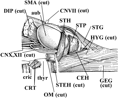
Fig. 11.
Ptilocercus lowii(Mammalia, Scandentia): ventral view of the musculature of the hyoid region of the right side of the body; muscles such as the geniohyoideus, sternothyroideus and thyrohyoideus are not shown (modified from Le Gros Clark, 1926 and Saban, 1968; the nomenclature of the structures illustrated basically follows that used in the present work; anterior is to the right). aub, auditory bulla; CEH, ceratohyoideus; CNVII, X, XII, cranial nerves VII, X and XII; cric, cricoid cartilage; CRT, cricothyroideus; DIP, digastricus posterior; GEG, genioglossus; HYG, hyoglossus; OM, omohyoideus; SMA, sternomastoideus; STEH, sternohyoideus; STG, styloglossus; STH, stylohyoideus; STP, stylopharyngeus; thyr, thyroid cartilage.

