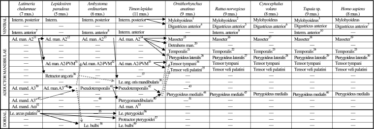
Table 1.
Scheme illustrating the authors’ hypotheses regarding the homologies of the mandibular muscles of adults of representative sarcopterygian taxa. The nomenclature of the muscles follows that used in the text; in order to facilitate comparisons, in some cases names often used by other authors to designate a certain muscle/bundle are given, between round brackets, in the text below the table; additional comments are given between square brackets. Data from evidence provided by our own dissections and comparisons and by a review of the literature (see text and Figs 3–17). Data from evidence provided by our own dissections and comparisons and by a review of the literature. The black arrows indicate the hypotheses that are most strongly supported by the evidence available; the grey arrows indicate alternative hypotheses that are supported by some of the data, but overall they are not as strongly supported by the evidence available as are the hypotheses indicated by black arrows (e.g. the overall analysis of the data available indicates that the urodele levator bulbi is a dorsal mandibular muscle, but the possibility that it derives from the adductor mandibulae cannot be completely ruled out: see text, Table 1, and Figs 3–17; VENTRAL, DORSAL = Ventral musculature and dorsal constrictor musculature sensuEdgeworth, 1935; ad. = adductor; ang. = anguli; interm. = intermandibularis; le. = levator; man. = mandibulae; mus. = muscles)
1[as described by e.g. Lightoller 1942, there is a mylohyoideus profundus, a mylohyoideus superficialis and, superficially to the latter, a digastricus anterior; Saban, 1971, states that these three structures come from the same embryological structure, i.e. that they seem to correspond to the intermandibularis posterior of other vertebrates: this is also supported by e.g. Jarvik 1963, 1980]
2[the mylohyoideus and digastricus anterior of rats clearly seem to correspond to the posterior, not the anterior, intermandibularis of other sarcopterygians, because the transversus mandibularis of rats corresponds to the intermandibularis anterior of other sarcopterygians; this is also supported by e.g. Bryant 1945]
3(posterior part of mylohyoid sensuLe Gros Clark 1924)
4[the correspondence between the mammalian digastricus anterior and part of the intermandibularis of other sarcopterygians is strongly corroborated by e.g. innervation (the intermandibularis and digastricus anterior are usually innervated by the ramus ventralis of CN5), ontogeny (e.g. the development of the marsupial Dasyrurus), and comparative anatomy of adults: see e.g. Edgeworth 1935]
5 (anterior belly of digastricus sensuGreene 1935)
6 (part of biventer sensuLeche 1886)
7 (anterior belly of biventer mandibulae sensuHuber 1930a)
8 (submentalis sensuIordansky 1992)
9 (transversus mandibularis sensuGreene 1935)
10 (anterior part of mylohyoid sensuLe Gros Clark 1924, and Sprague 1944a)
11 (adductor mandidulae ‘superficiel’sensuMillot & Anthony 1958)
12 (part of adductor mandidulae posterior sensuBemis & Lauder 1986)
13 (adductor mandibulae externus sensuIordansky 1992)
14 (adductor mandibulae externus sensuAbdala and Moro 2003)
15 (corresponds to the masseter + zygomatico-mandibularis, and possibly to the maxillo-mandibularis, sensuSaban 1971) [as shown in e.g. Saban's 1971 fig. 569, in the platypus specimens dissected by us the masseter is mainly divided into a deep part with anterior and posterior bundles and a superficial part with anterior and posterior bundles]
16[as described by Greene 1935, in the Norwegian rats dissected by us the masseter is mainly divided into a deep part with anterior and posterior bundles and a superficial part with anterior and posterior bundles]
17 (masseter + zygomatico-mandibularis sensuStafford & Szalay 2000) [in the colugo specimens dissected by us the masseter is subdivided into a superficial bundle, a deep bundle, and a zygomatico-mandibular bundle; the latter is sometimes considered as an independent muscle, but at least in the case of Cynocephalus, it is deeply mixed with the other masseter bundles]
18[as described by e.g. Le Gros Clark, 1924, in the Tupaia specimens dissected by us the masseter is mainly divided into deep, intermediate and superficial bundles]
19[in modern humans the masseter is usually mainly divided into deep and superficial bundles]11
20[some authors consider that the detrahens mandibulae is homologous to the digastricus anterior of other mammals, but this does not seem to be the case: see Saban 1968, p. 264; as stressed by e.g. Saban 1971, the detrahens mandibulae clearly seems to correspond to part of the adductor mandibulae A2 of non-mammalian tetrapods]
21[corresponds to part of the A2 of non-mammalian tetrapods but may possibly also include part of other adductor mandibulae structures such as the pseudotemporalis: see Barghusen 1968]
22[Greene 1935, describes the temporalis of rats as an undivided muscle, but as stated by Walker and Homberger, 1997, in the specimens dissected by us this muscle is divided into two bundles, one more superficial and anterior and the other more deep and posterior]
23[in the Cynocephalus specimens dissected by us the temporalis is not clearly divided into superficial and deep bundles, and there is no distinct pars suprazygomatica such as that found in Tupaia]
24[in the Tupaia specimens dissected by us the temporalis is mainly divided into a superficial bundle, a deep bundle, and a pars suprazygomatica sensuSaban 1971]
25[the temporalis of modern humans is usually described as an undivided muscle, but various authors, as e.g. Gorniak 1985, consider that it is in fact often divided into superficial and deep bundles]
26[in some parts of Edgeworth's 1935 work he seems to suggest that the pterygoideus lateralis and medialis are both included in the ’pterygoideus medialis’ of monotremes and that the pterygoideus lateralis only becomes separated in other extant mammals; however in other parts of Edgeworth's 1935 work he clearly states that the pterygoideus lateralis corresponds to part of the adductor mandibulae externus (= A2) of reptiles; more recent works, e.g. Barghusen 1968 and Jouffroy 1971, support this latter hypothesis; developmental data also indicate that the pterygoideus lateralis and pterygoideus medialis do not develop from the same anlage (e.g. Smith 1994); the platypus specimens dissected by us have both a pterygoideus lateralis and a pterygoideus medialis]
27 (pterygoideus externus sensuGreene 1935) [in the Norwegian rats dissected by us the pterygoideus lateralis is constituted by a single bundle]
28[in the Cynocephalus specimens dissected by us the pterygoideus lateralis is constituted by a single bundle]
29 (pterygoideus externus sensu Le Gros Clark 1924, 1926) [as described by e.g. Le Gros Clark 1924, in the Tupaia specimens dissected by us the pterygoideus lateralis is constituted by a single bundle]
30[in modern humans the pterygoideus lateralis is usually divided into superior and inferior heads: see e.g. Birou et al. 1991; Aziz et al. 1998; El Haddioui et al. 2005]
31 (part of adductor mandidulae posterior sensuBemis & Lauder 1986)
32 (adductor mandibulae posterior sensuIordansky 1992; levator mandibulae posterior sensuEdgeworth 1935 and Piatt 1938) [authors such as Piatt 1938 suggest that the A2-PVM of tetrapods as e.g. urodeles derives ontogenetically from the A3’ and/or A3’’, but the developmental work of Ericsson and Olsson 2004 strongly supports that it derives instead from the A2, as suggested by Diogo 2007, 2008 and Diogo et al. 2008]
33 (adductor mandibulae posterior sensuAbdala and Moro, 2003, and Holliday and Witmer, 2007)
34[there is some confusion regarding the origin of the tensor tympani and the tensor veli palatini; authors such as Brocks 1938, Barghusen 1986, and Smith 1992, state that it comes from the ‘pterygoideus posterior’ of reptiles; according to Edgeworth 1935, and Saban 1971 the mammalian tensor tympani and tensor veli palatini clearly correspond to the levator mandibulae posterior (= A2-PVM) of reptiles; our dissections and comparisons strongly support this latter view]
35[as described by e.g. Saban 1971, in the platypus specimens dissected by us the tensor veli palatini is present as an independent muscle]
36[seemingly derived from lateral portion of adductor mandibulae: e.g. Diogo 2007, 2008]
37[seemingly absent, but see 38]
38 (levator anguli oris sensuDiogo 2007, 2008) [present, somewhat mixed with A2; it may correspond to, or be derived/modified from, the retractor anguli oris of other sarcopterygians; we use the name ‘mandibularis’ to distinguish this muscle from the levator anguli oris facialis of certain mammals, which is a facial (hyoid), and not a mandibular, muscle]
39 (adductor mandidulae ‘moyen’sensuMillot and Anthony 1958)
40 (adductor mandidulae anterior sensuBemis & Lauder 1986)
41 (pseudotemporalis posterior and anterior sensuIordansky 1992; superficial and deep levator mandibulae anterior sensuEdgeworth 1935, and Piatt 1938; adductor mandibulae A3’ and A3’’sensuDiogo 2007, 2008)
42 (pseudotemporalis superficialis and profundus sensuAbdala and Moro, 2003, and Holliday and Witmer, 2007; adductor mandibulae A3’ and A3’’sensuDiogo, 2007, 2008)
43[the pseudotemporalis of non-mammalian tetrapods seems to correspond to part of the pterygoideus medialis, and possibly also to part of the temporalis, of extant mammals: see 21]
44[the pterygoideus medialis seems to correspond to the pseudotemporalis of amphibians such as Ambystoma, and, thus, to both the pseudotemporalis and pterygomandibularis of some other urodeles and some caecilians and of reptiles such as Timon: see also 49, 50, 51]
45 (pterygoideus internus sensuGreene 1935)
46 (pterygoideus internus sensuLe Gros Clark 1924, 1926)
47 (adductor mandidulae ‘profond’sensuMillot and Anthony 1958)
48[both the adductor A3’ and A3’’ seem to be included in the pseudotemporalis and/or pterygomandibularis of extant amphibians and reptiles: see Diogo 2007, 2008]
49[at least some caecilian and urodele amphibians have an independent muscle ‘pterygoideus’, which, according to Kleinteich and Haas 2007, probably corresponds to the pterygomandibularis of reptiles; in the Ambystoma ordinarium specimens dissected by us this ’pterygoideus’ is poorly differentiated from the pseudotemporalis]
50 (pterygoideus sensuHolliday & Witmer 2007) [seemingly derived from mesial portion of adductor mandibulae]
51[the pterygomandibularis of reptiles such as Timon seems to correspond to part of the pterygoideus medialis, and possibly also to part of the tensor tympani and/or tensor veli palatini, of extant mammals: see 34]
52 (intramandibular adductor sensuLauder 1980b)
53[in Timon the adductor mandibulae has a large and distinct anteroventral division that is lodged in the ‘adductor fossa’ of Lauder 1980b, and that is very similar to the Aω of other osteichthyans; similar adductor mandibulae structures are also found in other reptiles such as crocodilians, turtles and Aves: Edgeworth 1935; Holliday & Witmer 2007; according to Iordansky 2008, at least some of these ’Aω’ structures were acquired independently in evolution]
54[Edgeworth 1935 suggested that the dorsal mandibular musculature was probably acquired independently within gnathostomes, but the presence of this musculature is very likely plesiomorphic for this group, and perhaps for vertebrates as a whole: e.g. Holland et al. 1993; Diogo 2007, 2008]
55[the only dorsal mandibular muscle present in urodeles such as Ambystoma is the levator bulbi; amphibians such as caecilians have a ’levator quadrati’: see e.g. Kleinteich & Haas 2007; according to authors such as Edgeworth 1935, this latter muscle is derived from the adductor mandibulae, but authors such as Brocks 1938 argue that it is a dorsal mandibular muscle]
56[it is derived from the constrictor dorsalis, so it probably corresponds to part of the levator arcus palatini of Latimeria: Brocks 1938; Holliday & Witmer 2007; Diogo 2007, 2008]
57[it is derived from the constrictor dorsalis, so it probably corresponds to part of the levator arcus palatini or of e.g. Latimeria: see e.g. Brocks 1938; Holliday & Witmer 2007; Diogo 2007, 2008]
58[according to e.g. Edgeworth 1935 this muscle is derived from the adductor mandibulae; however, our dissections and comparisons support Brocks’ 1938 hypothesis, i.e. that the levator bulbi, as well as the ’levator quadrati’ of caecilians, are the remains of the constrictor dorsalis group in amphibians; according to Brocks 1938 the constrictor dorsalis group is conserved in many reptiles because of their kinetic skull]
59 (the levator bulbi sensu Frazzeta 1962, Haas 1997, and Schumacher 1973 seemingly corresponds to the tensor periorbitae sensuHolliday & Witmer 2007)

