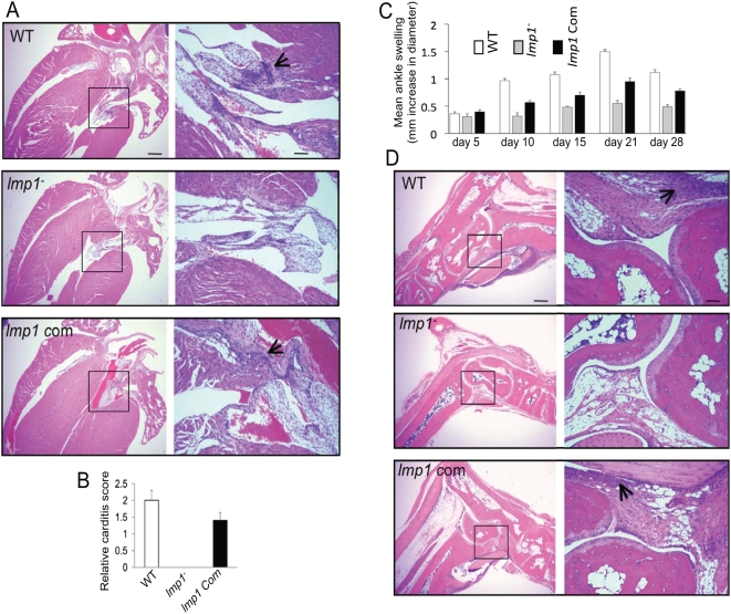
Figure 4. Deficiency of B. burgdorferi lmp1 reduces the severity of carditis and arthritis in mice.
(A) Representative histology of hearts isolated from mice infected with wild type B. burgdorferi (WT), lmp1 mutant (lmp1−), or lmp1 mutants complemented with lmp1 gene (lmp1 Com) isolates analyzed two weeks following infection. The left panel indicates lower-resolution (4×, bar = 400 µm) and higher-resolution (20×, bar = 80 µm) images of selected areas from corresponding sections (marked by box) shown in right panels. (B) Quantitative representation of the data shown in Figure 4A. At least ten random sections from each spirochete group were scored for severity of carditis in blinded fashion on a scale of 0–3 as indicated in Materials and Methods. (C) Severity of joint swelling in B. burgdorferi–infected mice. Arthritis was evaluated by assessment of the development of joint swelling of the mice infected with wild type B. burgdorferi (white bar), lmp1 mutant (gray bar), and lmp1 mutants complemented with lmp1 gene (black bar) isolates, measured using a digital caliper on day 5, 10, 15, 21, and 28 following spirochetes challenge. Bars represent the mean±SEM from three independent infection experiments. Differences in the joint swelling between groups of mice infected with lmp1 mutant and those with the lmp1-complemented isolates were significant (P<0.003) at all time points, except for day 5. (D) Representative demonstration of joint histology in mice infected with wild type or genetically manipulated B. burgdorferi isolates. Three weeks following spirochete infection, tibiotarsal joints were analyzed for histopathology. Both lower-resolution (4×, bar = 400 µm, left panel) and corresponding higher-resolution sections (20×, bar = 80 µm, marked by box, right panel) are shown. The arrows indicate the infiltration of inflammatory cells.

