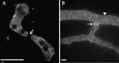
Figure 4.—
(A) Deconvolution microscopy of germling fusion and localization of PRM1-GFP in ΔPrm1 germling pairs [his-3∷Prm1-gfp; ΔPrm1∷hph (A24)]. PRM1-GFP showed vacuolar/endomembrane and some plasma membrane localization, but lacked localization to the fusion tips (arrow). (B) Strain A24 grown on MM agar containing sodium acetate as the sole carbon source for 4 hr. Examination of hyphae by deconvolution microscopy showed membrane localized GFP fluorescence (arrowhead) with higher intensity GFP fluorescence at points of hyphal fusion (arrow). Bars, 5 μm.

