Abstract
During development individual cells in tissues undergo complex cell-shape changes to drive the morphogenetic movements required to form tissues. Cell shape is determined by the cytoskeleton and cell-shape changes critically depend on a tight spatial and temporal control of cytoskeletal behavior. We have used the formation of the salivary glands in the Drosophila embryo, a process of tubulogenesis, as an assay for identifying factors that impinge on cell shape and the cytoskeleton. To this end we have performed a gain-of-function screen in the salivary glands, using a collection of fly lines carrying EP-element insertions that allow the overexpression of downstream-located genes using the UAS-Gal4 system. We used a salivary-gland-specific fork head-Gal4 line to restrict expression to the salivary glands, in combination with reporters of cell shape and the cytoskeleton. We identified a number of genes known to affect salivary gland formation, confirming the effectiveness of the screen. In addition, we found many genes not implicated previously in this process, some having known functions in other tissues. We report the initial characterization of a subset of genes, including chickadee, rhomboid1, egalitarian, bitesize, and capricious, through comparison of gain- and loss-of-function phenotypes.
DURING development and organogenesis most tissues arise from layers of epithelial cells that reorganize through complex morphogenetic movements. Many adult organs consist of tubular arrangements of epithelial sheets, and these tubules form during development through a process called tubulogenesis. There are a number of ways to generate tubules (Lubarsky and Krasnow 2003). One important process is the direct conversion of epithelial sheets into tubules through wrapping (Colas and Schoenwolf 2001) or budding (Hogan and Kolodziej 2002). Cells undergoing tubulogenesis change their shapes drastically from a cuboidal or columnar epithelial shape to a wedge shape or conical shape and then back to a more columnar epithelial shape once positioned inside the tube. Cell shape is determined by the intracellular cytoskeleton, primarily actin and microtubules. The cytoskeleton is closely coupled to cell–cell adhesion as well as to adhesion to the extracellular matrix. We are interested in understanding how the cytoskeleton and thus cell shape is regulated and coordinated during tubulogenesis.
We chose to perform a gain-of-function screen rather than a mutagenesis-based loss-of-function screen as phenotypes observed in the latter might be subtle and thus missed or phenotypes in a given tissue might be obscured by disruption of other tissues and many genes might also have redundant functions. In contrast, the gain-of-function/overexpression approach allows a particular tissue and gene to be targeted, and many such screens have been successfully conducted in the past (for examples, see Rørth et al. 1998; Molnar et al. 2006; Bejarano et al. 2008). The screen presented here uses the formation of the salivary glands in the Drosophila embryo as an assay system. The screen is based on a collection of transposable elements (EP elements) generated by Rørth et al. (1998) that contain UAS sites that respond to the yeast transcription factor Gal4 that is followed by a promoter directing expression, when activated, of genes located downstream 3′ of the EP insertion site. If combined through crosses with a tissue-specific source of Gal4 (Henderson and Andrew 2000; Zhou et al. 2001), overexpression (and in some cases antisense expression) of a downstream gene will be activated only in the target tissue, which in our case are the embryonic salivary glands in the Drosophila embryo.
Salivary gland formation in Drosophila is probably the simplest form of tubulogenesis (Lubarsky and Krasnow 2003). A patch of ∼200 cells in the ventral epidermis of the embryo within parasegment 2 becomes specialized to form a salivary gland primordium, the placode, with 100 cells on either side of the embryo. This fate determination occurs through a combination of the activities of the homeotic genes sex combs reduced (scr), extradenticle (exd), and homothorax (hth) and dorsal signaling by decapentaplegic (dpp) (Panzer et al. 1992; Henderson et al. 1999; Henderson and Andrew 2000). Without scr, exd, and hth function, no salivary glands form. Different subpopulations of cells are found in the invaginated gland, such as the secretory cells and the common and individual duct cells. Their distinction depends on EGF signaling from the ventral midline (Kuo et al. 1996; Haberman et al. 2003). Once the cells have become specialized at stage 10 of embryogenesis, no further cell division occurs within the primordium, and no cells are lost through apoptosis (Campos-Ortega and Hartenstein 1985; Bate and Martinez Arias 1993; Myat and Andrew 2000a). Invagination initiates in the dorsal posterior corner of the primordium, with all future secretory cells invaginating in a precise order, followed by invagination of the duct cells and formation of the ducts (Myat and Andrew 2000b). A key gene essential for the invagination is fork head (fkh). Fkh is a winged-helix transcription factor, and in its absence all of the cells fated to form the glands remain on the surface of the embryo as they fail to undergo apical constriction (Myat and Andrew 2000a). Once inside the embryo, the glands have to navigate their way through the surrounding tissues, including the visceral mesoderm and central nervous system, to reach their extended final position parallel to the midline and anterior–posterior axis. They are guided by cues from the surrounding tissues (Kolesnikov and Beckendorf 2005; Harris and Beckendorf 2007; Harris et al. 2007). Also, after initially invaginating in a posterior–dorsal direction, the glands turn and further extend into the embryo in a direction parallel to the anterior–posterior embryonic axis in a process dependent on integrins and downstream signals (Bradley et al. 2003; Vining et al. 2005).
A few factors that impinge on the cytoskeleton and cell shape during salivary gland morphogenesis have previously been identified. The actin cytoskeleton is modified through proteins such as Btk29/Tec29 in conjunction with Chickadee (Chandrasekaran and Beckendorf 2005). Small GTPases such as Rac and Rho affect the invagination of the glands (Pirraglia et al. 2006; Xu et al. 2008). Crumbs and Klarsicht affect the delivery of apical membrane and thus cell shape at late stages of morphogenesis (Myat and Andrew 2002). Nonetheless, how these factors work together throughout the whole process of invagination is still not clear, and it is likely that many others remain to be identified.
We have performed a gain-of-function/overexpression screen in the salivary glands with the aim of, first, identifying more genes that are required for salivary gland tubulogenesis (and thus potentially also for tubulogenesis in general) and, second, using this system as an assay for factors affecting the cytoskeleton and thus cell shape in general. The first aim assumes that genes that have a function in the morphogenesis of the glands and are endogenously expressed in the glands might perturb their invagination if overexpressed and if levels of expression are important. The second aim hypothesizes that overexpression of genes not endogenously expressed in the glands but important for cell-shape coordination in other tissues will lead to identifiable phenotypes in this screen, as defects in cell-shape changes resulting from the overexpression will affect the proper invagination of the glands. The orientation of some of the EP elements is also likely to lead to (over)expression of an antisense RNA, thus potentially inducing a tissue-specific loss-of-function effect. We identified seven genes that have previously been implicated in salivary gland morphogenesis or function, confirming the effectiveness of the screen, and also 44 insertions that uncover genes with potentially novel roles in the salivary glands or functions in the regulation of cell shape and the cytoskeleton in other tissues. Of these genes, 14 are previously uncharacterized genes. A selection of the genes that fall into the three categories discussed above (i.e., overexpression of a gene with a function in the glands; overexpression of a gene not expressed in the glands, revealing a function in cell-shape coordination in other tissues; and loss of function of a gene through tissue-specific antisense RNA expression) and recovered in the screen is examined in more detail below, including bitesize, egalitarian, chickadee, capricious, and rhomboid1.
MATERIALS AND METHODS
Screen design and fly husbandry:
fkhGal4 (Henderson and Andrew 2000; Zhou et al. 2001) was recombined on the third chromosome with a UAS construct containing GFP fused to the N terminus of the EF-Gas2 region of Shot (Subramanian et al. 2003) or the membrane-targeting domain of src fused to GFP (Kaltschmidt et al. 2000). One or the other of these marker lines were crossed to 1001 EP lines from the Rørth collection obtained from the stock centers in Szeged (second and third chromosomes; http://expbio.bio.u-szeged.hu/fly/index.php) and Bloomington (third chromosome; http://flystocks.bio.indiana.edu/). The fkhGal4 insertion was a gift from Deborah Andrew, the SrcGFP from Nick Brown. UAS-chickadee was from Lynn Cooley. rho[PΔ5], argos[lΔ7], flb[ik35], UAS-argos, UAS-CA-EGFR, UAS-sspi, UAS-caps, caps[PB1], trn[28.4], caps[Del1] trn[28.4] alleles were gifts from Matthew Freeman; egl[3e], egl[PR29], BicD[HA40], b BicD[18a], dp b Df(2L)TW119 and UAS-egl were gifts from Simon Bullock (the UAS-egl transgene leads to an approximately threefold increase in levels; S. Bullock, personal communication). To analyze egl mutant embryos, egl[3e]/egl[WU50] females were mated to egl[PR29]/+ males, and to analyze BicD mutant embryos, BicD[HA40]/+; b BicD[18a]/dp b Df(2L)TW119 mothers were mated to BicD[18a]/CyO males. In the detailed analyses (apart from the cases of egl and BicD mutant embryos), mutant embryos were identified by the absence of green balancer (balancer chromosomes used were CyO Kr∷GFP, TM3 Sb Ser twi-gal4 UAS-2x eGFP and TM6b Tb Sb df-Gal4 UAS-YFP). All other stocks used were from the Bloomington Stock Center. Crosses were maintained at 25° on cornmeal food, and embryos were collected on apple or grape-juice–agar plates.
EP lines were determined to be heterozygous or homozygous for the EP insertion. In the absence of visible balancer chromosomes, lines were assumed to be homozygous. Homozygous lines were crossed to a homozygous driver line, and embryos were collected overnight on apple or grape-juice plates with yeast paste. Twenty embryos between stages 10 and 13 and 20 embryos between stages 13 and 15 (an “early” and a “late” sample) were scored in live mounts in halocarbon oil (Halocarbon Oil 27, Sigma) after dechorionation in 50% bleach. In heterozygous balanced lines, 40 embryos in each of the early and late groups were scored, assuming equal fertilization and survival from both genotypes through the end of embryogenesis. Lines with ≥20% salivary gland defects, or with potential defects that would require quantitative analysis (i.e., changes in length), were subjected to second-pass screening. In the second pass, embryos were collected as above, fixed in 2:1 heptane:4% formaldehyde in PBS and stained with rhodamine phalloidin. A larger number of embryos were scored from these collections (average >80). First-pass hits were also crossed to w f flies, and embryos were collected and stained with rhodamine phalloidin as above to check for dominant positional effects of the EP insertion.
Immunohistochemistry, wide-field fluorescence, and confocal analysis:
Embryos were collected on grape-juice plates and processed for immunofluorescence using standard procedures. Briefly, embryos were dechorionated in 50% bleach, fixed in 4% formaldehyde, and stained with phalloidin or primary and secondary antibodies in PBT (PBS plus 0.5% bovine serum albumin and 0.3% Triton X-100). Crumbs and DE-Cadherin antibodies were obtained from the Developmental Studies Hybridoma Bank at the University of Iowa; the Shot antibody was raised in our lab and is identical in design to the one described in Strumpf and Volk (1998); the anti-dCrebA antibody was from Deborah Andrew (Andrew et al. 1997); the antiphosphohistone H3 and anti-GFP antibodies were from Abcam. Secondary antibodies used were Alexa Fluor 488 coupled (Molecular Probes) and Cy3 and Cy5 coupled (Jackson ImmunoResearch Laboratories), and rhodamine–phalloidin was from Molecular Probes. Samples were embedded in Vectashield (Vector Laboratories). Wide-field fluorescence images documenting the screen results were obtained on a Leica DMR (equipped with a MicroFire camera, Optronics), a Zeiss Axioplan 2 (equipped with a Princeton Instruments camera), and a Zeiss Axioskop Mot 2 (equipped with a Jenoptik C14 camera), using PictureFrame, Metamorph, and Openlab software, respectively. Confocal images were obtained using an Olympus Fluoview 1000. Confocal laser, iris, and amplification settings in experiments comparing intensities of labeling were set to identical values. Wide-field fluorescence and confocal images were assembled in Adobe Photoshop, and confocal z-stacks and z-stack projections were assembled in Image J.
In situ hybridization:
In situ hybridization of whole-mount embryos was performed essentially as described by Tautz and Pfeifle 1989). To combine the in situ protocol with immunohistochemistry for GFP, the anti-GFP antibody was incubated together with the anti-DIG antibody, followed by fluorescent secondary antibody incubation to reveal the GFP after the BCIP/NBT color reaction. Images were obtained on a Leica DMR (equipped with a MicroFire camera, Optronics) and composites were assembled using Adobe Photoshop.
The following primers were used to generate in situ probes: chic 5′-TTTCCATCTACGAGGATCCC, chic 3′-ATTTCGTTCAAAGCTGAGGAC; caps 5′-CGGGCAATTACCATGTCGTTG, caps 3′-GATGTGGCTGATGCGATTCTG; trn 5′-GTGGGCATCTGGTGCATTTTG, and trn 3′-GATAAAGGATGCGCAACTGGG. For the btsz probe, the cDNA clone AY229970 was used to transcribe antisense and sense probes.
Statistics:
We determined the base rate of salivary gland defects observable in our experimental stocks by counting defects in the genotype +/+; +/CyO; fkhGal4∷UAS-GFPmarker/+. The base rate in this genetic background was determined as 4.3% (n = 748). A similar base rate was obtained in a genetic background where the CyO balancer chromosome was replaced by the GFP-marked chromosome +/+;+/btlGal4∷UAS-GFP; fkhGal4∷UAS-GFPmarker/+; the base rate was 4.4% (n = 878). Thus, we exclude any effect at least of a CyO balance chromosome present on salivary gland morphogenesis.
To set a cutoff level for the rate of affected salivary glands counted in each experiment, above which we determined that EP elements driven by fkhGal4 affect the salivary gland morphogenesis, we chose an arbitrary 20% defects as a cutoff for the first-pass analysis. This yielded 187 EP lines (18.6% of the total lines screened) to be rescreened in the second-pass analysis, yielding 51 confirmed insertions overall. This equals ∼5% of the total number of EP lines screened, which also equals 2 SD from the mean of a normal distributed sample, indicating that we set our cutoff at a sensible level.
RESULTS
Experimental design of the gain-of-function screen:
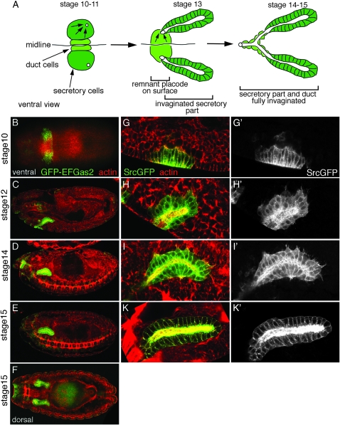
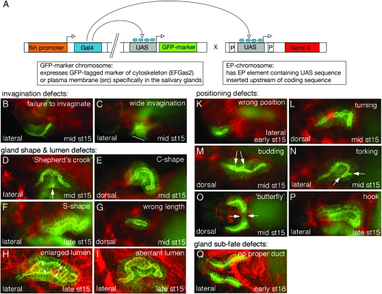
To address how the cytoskeleton and cell shape is regulated during such a process of tubulogenesis, we performed a gain-of-function screen in the salivary glands of the Drosophila embryo. We used a gland-specific Gal4-driver, fkhGal4 (Henderson and Andrew 2000; Zhou et al. 2001), to drive expression of either a marker of the microtubule cytoskeleton, GFP-EFGas2 (Subramanian et al. 2003), or a marker of cell shape, SrcGFP (Kaltschmidt et al. 2000), in the glands only. Flies carrying these marker chromosomes (GFP-EFGas2 or SrcGFP marker plus fkhGal4: marker line) were crossed to a collection of EP-element lines containing UAS elements, leading to the expression of gene X located 3′ downstream from the EP-element insertion site. We drove expression from 1001 EP elements specifically in the salivary glands and screened for any apparent problems in their morphogenesis (see Figure 1 for wild-type morphogenesis and marker expression and Figure 2A for a scheme explaining the screen setup). It has previously been shown that the proper invagination and positioning of the salivary glands depends on the surrounding tissues such as the visceral mesoderm (Vining et al. 2005). The tissue-specific expression of genes in the screen allowed us to identify factors that acted within the glands themselves and did not affect functioning of the surrounding tissues, thus giving a phenotype due to a secondary defect.
Figure 1.—
Salivary gland development visualized using fkhGal4-driven GFP-marker expression. Salivary gland morphogenesis from embryonic stages 10–15. (A) Schematic of salivary gland invagination, ventral view. (B–F) Low-magnification confocal sections of embryos stained with phalloidin to reveal actin (red) and expressing GFP-EFGas2 under the control of fkhGal4 in the salivary glands (green). (C–E) Lateral views. (B) A ventral view. (F) A dorsal view. (G–K′) Close-up confocal sections of salivary glands labeled with phalloidin to reveal actin (red) and expressing SrcGFP under the control of fkhGal4 (green, and as a single channel in G′–K′). G–K show lateral views. Note that GFP-EFGas2 labels microtubules, whereas SrcGFP is targeted to the membrane and thus reveals cell shape.
Figure 2.—
Phenotypes observed upon overexpression of genes in the salivary glands using fkhGal4. (A) Schematic of the setup of the screen. The phenotypes observed in the screen could be classified according to the categories depicted in this figure. Broad categories are invagination defects (B and C), gland shape and lumen defects (D–I), positioning defects (K–P), and gland subfate defects (Q). (B–Q) GFP-marker expression in green and phalloidin staining to reveal actin in red. Lateral or dorsal views and embryonic stages of embryos are indicated in each panel. The line in C indicates the area of the too-wide opening of the invaginating gland shown; the arrow in D points to where proximal and distal cells of the gland touch due to excessive bending; the double arrows in H indicate the too-wide width of the gland; the arrows in M point to two buds emerging from the side of the gland; the arrows in N point to the two ends of a fork; the arrows in O point to cells of the glands that appear to touch across the midline. B–G and K–Q are wide-field fluorescence images; H, I, and Q are confocal sections.
We crossed flies of marker to flies carrying an EP insertion on the second or third chromosome (see scheme in Figure 2A). The resulting offspring overexpressed a gene X specifically in the salivary glands. These embryos were collected early (stages 10–13) and late (stages 13–15) during embryogenesis and analyzed live for any apparent defect in salivary gland invagination, positioning, and shape of salivary gland cells or gland lumen. When phenotypes were observed in >20% of embryos, embryos were collected again, fixed, and counterstained for actin using phalloidin to analyze general morphology. Positive insertions were defined as having 20–90% of embryos showing a salivary gland phenotype after the second examination. The baseline rate of obtaining salivary glands with a phenotype in embryos expressing GFP-EFGas2 or SrcGFP in the glands under the control of fkhGal4 was ∼4% (see materials and methods). In some cases the position and orientation of EP elements would be predicted to lead to the overexpression of an antisense RNA rather than a coding-sense mRNA. Positive insertions resulting from presumed antisense RNA expression are indicated as such in Table 1.
TABLE 1.
Genes identified in the gain-of-function screen
| EP no. | Cytology | Gene affected | Effect, location in gene, direction | Penetrance of phenotypesa | Phenotype in salivary glands (fkhGal4) | Function or mutant phenotype, known function in flies or glands |
|---|---|---|---|---|---|---|
| Cytoskeleton and cytoskeleton associated | ||||||
| EP(2)570 | 2L (26B4) | CG13993 (actin/tubulin protein folding) | Overexpression, 5′-end of gene | Strong | Variable | No mutant available |
| EP(2)713 | 2L (26B1) | chickadee (profilin) | Overexpression, in 5′ region of gene upstream of most CDS | Strong | Variable | Lethal, Tec29 chic double mutants show salivary gland phenotype (Chandrasekaran and Beckendorf 2005) |
| EP(2)1011 | 2L (26B1) | chickadee (profilin) | Antisense to 5′ 1 kb of gene (or overexpression of eIF4a, 1.8 kb downstream) | Strong | Hooks, shepherd's crook | Lethal, Tec29 chic double mutants show salivary gland phenotype (Chandrasekaran and Beckendorf 2005) |
| EP(2)938 | 2R (59F7) | egalitarian (dynactin associated) | Overexpression, directly 5′ of gene | Strong | Variable | Female sterile, lethal, microtubule-based transport |
| EP(2)2047 | 2R (57E5) | syndecan | Inserted in 5′-end of sdc wrong strand, could drive antisense to 6 of the 90-kb sdc locus or overexpress sara 7 kb downstream | Strong | Variable | Lethal, works in conjunction with Slit (Steigemann et al. 2004) |
| EP(3)3567 | 3R (88D5) | bitesize (synaptotagmin-like protein) | Middle of gene, could drive antisense to most isoforms | Weak | Variable | Lethal, actin organization at adherens junctions (Pilot et al. 2006) |
| Signaling | ||||||
| EP(2)578 | 2L (24E1) | traf-4 (TNF-receptor-associated factor 4, previously called Traf-1 in flies) | Overexpression, middle of gene, upstream of most CDS | Very strong | Fewer cells in glands, overexpression reportedly induces apoptosis (Kuranaga et al. 2002) | Larval lethal (Kuranaga et al. 2002) |
| EP(2)2167 | 2L (29A1) | btk29/tec29 (Btk family kinase) | 3′-end of gene wrong strand, could drive antisense to >30 of 40 kb btk29A locus | Weak | Variable | Lethal; Tec29 is important for salivary gland invagination (Chandrasekaran and Beckendorf 2005) |
| EP(2)1173 | 2L (37E1) | ranGAP | Overexpression, middle of gene but upstream of CDS | Strong | Hook, crook | Viable; possible link between nuclear transport, actin, and profilin (Minakhina et al. 2005) |
| EP(2)2158 | 2L (37D2) | doughnut on 2 (RYK family receptor tyrosine kinase) | Overexpression | Weak | Variable | Important for salivary gland positioning as is drl another RYK (Harris and Beckendorf 2007) |
| EP(3)3542 | 3L (61C1) | ptpmeg (tyrosine phosphatase) or mthl9 (G-protein-coupled receptor) | Overexpression of ptpmeg or antisense of mthl9 (intronic to ptpmeg) | Strong | Budding | Viable; Ptpmeg is FERM domain protein |
| EP(3)3704 | 3L (62A2) | rhomboid1 (EGF signaling, intramembrane protease) | Overexpression | Very strong | Aberrant duct morphogenesis, potentially due to overproliferation (see Figures 4 and 5) | Mutations in rho or spitz lead to transformation of duct cells into secretory cells (Kuo et al. 1996) |
| EP(2)2201 | 2L (37F2) | spitz (secreted EGF ligand) | Inserted into middle of spi; could drive antisense to 4 kb of some spi mRNAs or overexpress msb1l 5 kb downstream | Weak | Variable | Mutations in rho or spitz lead to transformation of duct cells into secretory cells (Kuo et al. 1996) |
| EP(3)549 | 3L (62E7) | misshapen (Ste20 kinase) | Overexpression, directly 5′ of gene | Weak | Too-wide and lumpy lumen | Lethal, linked to nuclear movement via BicD (Houalla et al. 2005) and cell-shape changes during morphogenesis (Koppen et al. 2006) |
| Nucleus, transcription (factors) | ||||||
| EP(2)2176 | 2R (48E4) | Smd3 (snRNP, splicing) | Overexpression, 5′ of gene | Strong | Hooks | Lethal (Schenkel et al. 2002) |
| EP(2)474 | 2L (21B5) | kismet (helicase) | Overexpression, in 5′ region of gene upstream of all CDS | Weak | Too-wide lumen | Lethal, segment specification (Daubresse et al. 1999) |
| EP(2)993 | 2R (50E1) | combgap (zinc-finger protein) | Inserted into 5′-end of CG30096 wrong strand, antisense to cg 2 kb away | Very strong | Variable | Lethal; hedgehog signaling in leg patterning (Svendsen et al. 2000) |
| EP(3)486 | 3L (75E1) | ftz-f1 (ftz-transcription factor1) | Inserted into locus, should overexpress longer isoform | Weak | Variable | Lethal (Florence et al. 1997) |
| EP(3)711 | 3L (64E8) | bre-1 (nuclear factor downstream of Notch) | Overexpression, directly 5′ of gene | Weak | Variable | Lethal (Bray et al. 2005) |
| Protein synthesis and degradation | ||||||
| EP(2)463 | 2R (47F7) | Tapδ (translocon-associated protein δ) | Overexpression, 5′-end of gene | Weak | Variable | Lethal, downstream of dCreb-A in the glands (Abrams and Andrew 2005) |
| EP(2)2063 | 2L (37B7) | nedd8 (regulation of proteolysis) | Overexpression, 5′-end of gene | Weak | Butterfly | Lethal, ubiquitin like, cooperates with cullin3 (Zhu et al. 2005) |
| EP(2)1187 | 2L (33C1) | CG5317 (ribosomal subunit) | Overexpression, inserted in 5′-end of JhI-21, wrong strand, should drive CG5317 400 bp downstream | Strong | Shepherd's crook | ND |
| Membrane traffic | ||||||
| EP(2)2028 | 2R (48F8) | garz (arf-GEF, GBF1) | Overexpression, 5′-end of gene | Weak | Severe hooks | ER-to-Golgi trafficking in mammals (Szul et al. 2007) |
| EP(2)2313 | 2L (35F1) | syntaxin5 (SNARE protein) | Overexpression, directly 5′ of gene | Weak | Degenerating glands | Lethal, membrane fusion, cytokinesis (Xu et al. 2002) |
| Cell surface and extracellular | ||||||
| EP(2)827 | 2R (58D4) | CG3624 (Ig domain protein) | Overexpression, directly 5′ of gene | Strong | Variable | ND |
| EP(2)937 | 2R (52D1) | slit (axon guidance receptor) | Middle of gene, wrong strand, antisense to 20 of 50 kb gene? | Strong | Hooks | Lethal; slit has been shown to be involved in salivary gland positioning (Kolesnikov and Beckendorf 2005) |
| EP(2)2120 | 2L (22A3) | CG14351 (LRR and Ig domain transmembrane protein) | Overexpression, directly 5′ of gene | Weak | Variable | ND; BLAST shows similarity to Slit |
| EP(2)2463 | 2L (35D4) | gliotactin (transmembrane protein of septate junctions) | Overexpression, directly 5′ of gene | Weak | Variable | Lethal; important for tube size control in trachae (Paul et al. 2003) |
| EP(3)552 | 3L (70A3) | capricious (transmembrane LRR protein) | Overexpression, directly 5′ of gene | Very strong | Bizarrely branching and budding lumen | Lethal (Shishido et al. 1998) |
| Enzymes | ||||||
| EP(2)2199 | 2R (51B1) | tout velu (glucosaminyl-transferase) | Inserted in intron of both ttv and lamC (which is intronic to ttv), could overexpress ∼10 of 60 kb ttv (∼50% of CDS) or 1.1 kb antisense to LamC | Weak | Variable | Lethal; mutants disrupt hh, wnt, and dpp signaling (Bornemann et al. 2004) |
| EP(2)1157 | 2R (59B6) | CG9849 (potential protease of the subtilase family) | Inserted into 5′-end of CG3800, wrong strand, overexpression of CG9849 600 bp away | Weak | Variable | ND |
| EP(3)3639 | 3L (65A10) | CG10163 (phospholipase A1) | Inserted 5′ of Best2, wrong strand, could drive antisense to CG10163 800 bp away | Weak | Too large and irregular lumen | ND |
| Mitosis, meiosis, and germline | ||||||
| EP(2)812 | 2L (35C1) | vasa or vig (vasa intronic gene) | In 3′ region of vig, which is intronic to vasa, would overexpress 3′ 1 kb of vig or antisense to vasa | Weak | Lumpy lumen | vasa: lethal, germ cell determination; vig: ND |
| EP(3)341 | 3R (82D2) | tacc (centrosomal protein) | Middle of gene, could drive antisense to all large isoforms of tacc | Weak | Variable | Lethal (Barros et al. 2005) |
| Other | ||||||
| EP(2)2356 | 2R (57A6) | mir-310/-313 cluster | Overexpression, inserted 200 bp 5′ of cluster | Very strong | Too-wide irregular lumen | Disruption of mir-310 cluster affects dorsal closure (Leaman et al. 2005) |
| EP(2)2586 | 2R (57A6) | mir-310/-313 cluster | Overexpression, inserted 100 bp 5′ of cluster | Strong | Irregular lumen | Disruption of mir-310 cluster affects dorsal closure (Leaman et al. 2005) |
| EP(2)2587 | 2R (57A6) | mir-310/-313 cluster | Overexpression, inserted 100 bp 5′ of cluster | Weak | Irregular lumen | Disruption of mir-310 cluster affects dorsal closure (Leaman et al. 2005) |
| EP(2)1221 | 2L (27F4) | mir-275, mir-305 | Overexpression, 2 kb upstream of genes | Strong | Shepherd's crook | ND |
| EP(2)2083 | 2R (45F1) | CG1888 | >6 kb away, overexpression | Weak | Variable | ND |
| EP(2)1163 | 2L (33E4) | vir-1 (virus-induced RNA 1) | Overexpression, directly 5′ of gene | Weak | Budding | ND |
| EP(2)1239 | 2L (25F5) | CG14005 or CG7239 | Inserted in 5′-end of CG9171 wrong strand, antisense to CG14005 300 bp downstream or overexpression of CG7239 2 kb downstream | Weak | Variable | ND; both CG14005 and CG7239 conserved only among Drosophilidae |
| EP(2)2219 | 2L (33E4) | CG6405 | Overexpression, inserted 1.5 kb upstream of CG6405 | Weak | Variable | ND; two mammalian orthologs |
| EP(2)2190 | 2R (55E1) | CG30332 | Overexpression, 1 kb 5′ of CG30332 | Strong | Hooks | ND |
| EP(2)2182 | 2R (54A2) | CR30234 (cytosolic tRNA gene) | Overexpression | Very strong | Variable | ND |
| EP(3)313 | 3R (98E5) | CG1523 (related to WD40 repeat-containing protein 32) | Overexpression, inserted directly 5′ of gene | Strong | Variable | ND |
| EP(2)2269 | 2R (53D11) | CG34460 | Antisense to CG34460?, EP is ∼2.5 kb away | Weak | Variable | ND |
| EP(2)383 | 2L (23C4) | Nothing downstream for >10 kb; next CG is CG17265 14 kb away | Inserted 3′ of CG3558 | Weak | Short, expanded at turn | ND |
| EP(2)2173 | 2L (35B2) | nothing downstream for >10kb | Inserted into 5′-end of no ocelli, wrong strand | Weak | Variable | ND |
| EP(2)2146a | Unclear | Unclear | Genome position of EP unclear | Strong | Searching cells | ND |
| EP(2)2265 | Unclear | Unclear | Genome position of EP unclear | Very strong | Short, straight, expanded | ND |
| EP(2)985 | Unclear | Unclear | Genome position of EP unclear | Strong | Budding, branching | ND |
All the genes identified in the screen, sorted according to their proposed function, are listed.
Penetrance of phenotypes: weak, 20–30% of embryos showing phenotype; strong, 30–50% of embryos showing phenotype; very strong, >50% of embryos showing phenotype (with three cases <70% and four cases >90%).
Phenotypes identified in the screen:
We screened 1001 EP-element insertions on the second and third chromosome (a list of all lines screened can be found in supplemental Table 1). Overexpression in the salivary glands of genes located downstream from EP elements under the control of fkhGal4 led to a variety of phenotypes that could be classified in four major classes (Figure 2): invagination defects (failure to invaginate, wide invagination; see Figure 2, B and C); gland shape and lumen defects (shepherd's crook, C-shape, S-shape, wrong length, enlarged lumen, aberrant lumen; see Figure 2, D–I); positioning defects (wrong position, turning, budding, forking, hook, butterfly; see Figure 2, K–P); and gland subfate defects (no proper duct; see Figure 2Q). These overall phenotypes suggest that the screen detected interference at all stages of salivary gland formation.
Although the phenotypes listed above were recurrently found in the screen, half of the positive insertions showed a variable phenotype, combining several of these individual phenotypes. The other half showed a consistent phenotype restricted to one class or even to one specific phenotype (see Table 1, Phenotype in salivary glands). This suggests that, in the cases of genes showing variable phenotypes upon overexpression in the glands, a specific phenotype was not necessarily reflective of only a certain process failing during the invagination, but rather indicated that many perturbances at the molecular level might lead to similar phenotypes.
Genes identified in the screen:
Of 1001 EP lines screened, 51 showed a phenotype in the salivary glands when crossed to fkhGal4, equalling 5.09% of the total lines analyzed. The penetrance of phenotypes varied from weak (2.7% of EPs tested) to strong (1.7% of EPs tested) to very strong (0.7% of EPs tested; see Table 1). The genes affected could be classified according to their predicted function (see Table 1). Several of the genes identified by positive insertions have previously been implicated in salivary gland morphogenesis or function within the glands: chickadee (Chandrasekaran and Beckendorf 2005), tec29 (Chandrasekaran and Beckendorf 2005), doughnut on 2 (Harris and Beckendorf 2007), rhomboid1 and spitz (Kuo et al. 1996), tapδ (Abrams and Andrew 2005), and slit (Kolesnikov and Beckendorf 2005). Three of the insertions identifying these could potentially induce antisense RNA expression and could thus mimic a loss-of-function situation (chic, tec29, slit), and three insertions could induce overexpression (dnt, rhomboid1, spitz). Overexpression of dnt could affect the positioning cues that the migrating glands receive, whereas rhomboid1 and spitz overexpression would lead to excess Spitz ligand being provided, potentially overstimulating EGFR signaling (see below). These genes served as confirmation that factors impinging on salivary gland morphogenesis were picked up in this screen.
The majority of positive insertions (44 of 51 EPs), however, were inserted into genes that have not previously been implicated in salivary gland morphogenesis or in tubulogenesis in general. Several of the encoded proteins have known functions in other tissues in flies such as Egalitarian (Navarro et al. 2004), Traf-4 (Cha et al. 2003), RanGAP (Kusano et al. 2001), Smd3 (Schenkel et al. 2002), Nedd 8 (Zhu et al. 2005), and Tout-velou (The et al. 1999). Fourteen of the 51 hits were EPs inserted in previously uncharacterized genes, many with close orthologs in other species, including vertebrates.
Analysis of individual genes in detail:
In the following section we will discuss a subset of the genes identified in the screen. Three of these were genes endogenously expressed in the glands (chickadee, rhomboid1, and egl), and thus overexpression could have interfered with their proper function in the glands. One gene (btsz) was expressed in the glands and was identified through an insertion that would have led to production of a tissue-specific antisense RNA, thus potentially mimicking a loss-of-function situation. The last gene (caps) was not endogenously expressed in the glands and thus the overexpression identified a potential requirement elsewhere for proper cell and tissue shape.
Genes endogenously expressed in glands identified through overexpression:
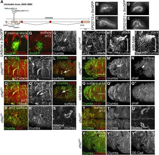
Chickadee encodes the Drosophila Profilin protein. Profilins are actin–monomer-sequestering proteins, which have been implicated in promoting both actin polymerization and depolymerization (Yarmola and Bubb 2006). Drosophila Profilin fulfills essential functions at all stages of development and also in the female germline (Verheyen and Cooley 1994). Loss of profilin has been associated with the inability to constrict apical surfaces in the morphogenetic furrow of the larval eye disc (Benlali et al. 2000). With respect to salivary gland morphogenesis, it has been reported that tec29 chic double mutants show disorganized actin in the salivary gland placode and display a delay in invagination (measured by the remaining placode area at stage 14). This study also reports that chic mutant embryos have normal glands (Chandrasekaran and Beckendorf 2005). Two EP insertions into chic showed phenotypes in the glands when driven by fkhGal4, EP(2)713, and EP(2)1011. EP(2)713 should overexpress the entire chickadee coding sequence (see supplemental Figure 1), whereas EP(2)1011 is inserted in the opposite direction and could drive expression of an antisense RNA to the 5′ most 1 kb of the chic pre-mRNA (or it could drive expression of eIF4a, situated 1.8 kb away; see scheme in Figure 3A). Expression of either EP led to aberrantly shaped glands (Figure 3, B–E), with EP(2)1011 giving frequent hook-shaped and shepherd's crook-shaped glands (Figure 3, D and E). Overexpression of a UAS-chickadee construct using fkhGal4 led to embryos showing a disorganized epidermis in the regions where fkhGal4 was expressed, with a loss of apical Crumbs accumulation in the epithelial cells of the epidermis (Figure 3, F–G′). The glands nonetheless invaginated and, within the invaginated portion of the glands, Crumbs was localized apically. This suggests that epithelial integrity and/or polarity might be impaired if levels of Profilin are imbalanced. Effects on junctional Armadillo in the absence of Profillin have been described (Townsley and Bienz 2000). In chic mutant embryos (either chic221 or chic01320) at a stage when the first cells had just invaginated from the salivary gland placode, cell shapes within the placode often appeared irregular compared to wild type, although Crumbs was still localized apically at this stage (compare H, H′, and I in Figure 3). At later stages the salivary glands invaginated but were irregular in shape, and the placodal and surrounding epidermal cells on the surface of the embryo appeared disrupted with absent or mislocalized Crumbs labeling (Figure 3, K and L and O and P at stage 12 and M and N at stage 14; for comparison, a matching wild-type epidermal scan at stage 14 is shown in Q–Q″). Other apical markers such as the spectraplakin Shot and DE-Cadherin also appeared disrupted at these stages (Figure 3, V–V″ for DE-Cadherin and data not shown). Nonetheless, within the invaginated portion of the glands Crumbs was localized apically, probably because early apical Crumbs localization in the placodal cells was unperturbed. Thus, in contrast to an earlier report (Chandrasekaran and Beckendorf 2005), either elevation or disruption of Chickadee/Profilin levels appeared to affect salivary gland invagination to some extent. As Profilin has been shown to promote both actin polymerization and depolymerization, depending on the context and tissue (Yarmola and Bubb 2006), an imbalance of Profilin levels (either decreased or increased) could affect the critical cortical function of actin during cell-shape changes required to allow the invagination and/or the cell rearrangements on the surface of the embryo during invagination.
Figure 3.—
Chickadee (Profilin) is important for salivary gland invagination. chickadee encodes the Drosophila Profilin protein. (A) Scheme of the chic locus indicating the position and orientation of the two EP lines that showed phenotypes when driven in the salivary glands. B and C and D and E show phenotypes observed in the screen for EP713 and EP1011 respectively; wide-field fluorescent images of live embryos are shown. (F–G′) Overexpression of chickadee using a UAS-chickadee construct led to invagination problems and aberrantly shaped glands. (F) An internal confocal stack of a gland (14 μm thick), labeled with Crumbs to reveal the apical surface/lumen of the glands (red) and showing the SrcGFP marker in green. (G and G′) A surface stack of the same embryo (with Crumbs in red and SrcGFP in green in G and Crumbs as a single channel in G′; 3 μm thick). The arrow G′points to disrupted epidermis in the region of the placode from which the glands have started to invaginate. Note the absence of Crumbs from the apical surface of cells in this region. (H and H′) A low and high magnification view, respectively, of the slightly disorganized epidermis of a chic mutant embryo labeled for crumbs, with H′ showing the salivary gland placode and gland in a projection (18-μm thick stack). (I) A wild-type placode and gland (17-μm thick stack) at the same stage as in H′. Note that the highly organized arrangement of apical constriction of the placodal cells is less apparent in the chic mutant (bracket in H′ and in I). K and L and O and P show the aberrant glands and disrupted epidermis in two different chic alleles (chic01320 and chic221) at stage 12; labeling for Crumbs is in green (and also shown as a single channel in K′ and O′) and for phalloidin in red. Note the disruption and absence of apical Crumbs labeling in the region of the salivary gland placode (arrows in K′, L, O′, and P point to these areas). (K) A projection of a 35-μm thick stack. (L) A 5-μm-thick surface projection. (O) A projection of a 26-μm-thick stack. (P) A 3-μm-thick surface projection. (M and N) The disrupted epidermis in the region from which salivary gland cells invaginated in a stage 14 embryo. Crumbs is green in M and a single channel in M′, and phalloidin is red in M and a single channel in N. M is a projection of a 34-μm-thick stack, and N is a 5-μm-thick surface stack. For comparison, a stage 14 wild-type embryo is shown in Q–Q″. Crumbs is green in Q and a single channel in Q′, and phalloidin is red in Q and a single channel in Q″; Q is a 5-μm-thick surface stack. The arrows in M′ and N point to the disrupted region, the white lines in M and Q indicate the ventral midline (the view in M–N is slightly oblique). (R–V″) chic221 mutant embryos at stages 12–14. R and R′ show lateral views of a placode, whereas S shows an internal stack of the gland. T and T′ are ventral views of the two placodes, with U showing an internal stack of the glands. (V–V″) A surface stack of a mutant embryo (5 μm thick). Crumbs is green in R, T, and V and a single channel in R′, T′, and V′; phalloidin is red. Both S and U show Crumbs labeling to outline the lumen of the gland. DE-Cadherin (DE-Cad) labeling is in red in V and as a single channel in V″.
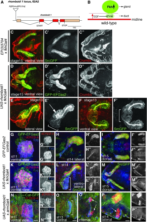
Rhomboid1 and EGF-receptor signaling are known to influence cell-fate decisions within the salivary gland primordium. The EGF-ligand Spitz is secreted from the ventral midline cells with Rhomboid being the intramembrane protease essential for its release (Shilo 2005). Spitz diffuses a few cell diameters laterally to induce the most ventral cells within the salivary gland placode to become duct cells, whereas the other placodal cells become secretory cells (see scheme in Figure 4A). This switch in fate transmitted by EGF is in part achieved through repression of the fkh transcription factor. Fkh in turn represses three duct-specific genes: trachealess (trh), dead ringer (dri), and serrate (ser) (Kuo et al. 1996; Haberman et al. 2003). Thus, by the end of embryogenesis, rhomboid1 and other spitz group mutants have salivary glands that are composed entirely of secretory cells and are completely enclosed within the embryo without any ductal connection to the outside (Figure 5, A–B′; Kuo et al. 1996).
Figure 4.—
rhomboid1 overexpression disrupts salivary gland morphogenesis, but is not sufficient to induce salivary duct fate. (A) Scheme of the rho locus indicating the position of the EP identified in the screen. (B) Scheme depicting the known involvement of EGF signaling in salivary gland morphogenesis. EGF is released from the midline (red line) and induces, in the cells close to the midline (light green), the repression of fkh, which in turn leads to suppression of secretory fate in these cells, inducing them to adopt duct cell fate. Fkh expression remains high in the remaining salivary gland primordium (dark green), thus inducing these cells to form the secretory part of the gland (Kuo et al. 1996). (C–C″) Overexpression of rhomboid1 in the salivary glands using EP(3)3704 led to glands that, at stage 15 of embryogenesis, were located too far anterior with secretory cells that appeared cuboidal instead of columnar, no proper duct connecting the secretory portions to the outside, and an aberrantly shaped lumen. The SrcGFP marker is green in C and a single channel in C′; phalloidin is red in C and a single channel in C″. (D–D″) The same phenotype as in C is observed when a UAS-rhomboid1 construct is expressed in the glands using fkhGal4. The GFP-EFGas2 marker is green in D and a single channel in D′; Crumbs is red in D and a single channel in D″. (E–F′) Already at stage 13 the invaginated portion of the gland shows aberrant morphology (ectopic lumen indicated by the arrow in E′), and the amount of cells remaining at the surface appears too large (bracket in E′ and F′). The SrcGFP marker is green in E and F (and a single channel in E′ and F′); crumbs is in red. (G–P″) Analysis of dCreb-A and Eyegone expression: markers of secretory and duct fate, respectively. (G–I″) Control glands expressing only the GFP-EFGas2 marker labeled with antibodies against dCreb-A and Eyg at stage 11 (G–G″), stage 14 (H–H″), and stage 15 (I–I″). (K–M″) Glands expressing UAS-rhomboid1 in the salivary glands using fkhGal4 labeled for dCreb-A and Eyg at stage 11 (K–K″), stage 14 (L–L″), and stage 15 (M–M″). Note that, despite the irregular shape and ectopic cells (bracket in L), dCreb-A is strongly expressed in the early invaginated part of the glands (L′ and M′). Eyg is expressed in the most anterior cells of the invaginated glands (L″ and M″), as in the control, and also in the ectopic cell bulge on the surface of the embryo (L″; bracket in L denotes the bulge, and the dashed line indicates the ventral midline). (N–P″) Glands expressing UAS-rhomboid1 using armGal4. (N–N″) dCreb-A and Eyg expression in the placode at stage 11. (O and O′) Ventral view of the remaining placode (O) and invaginated glands (O′) at stage 13. More ectopic cells expressing Eyg are found on the ventral surface (bracket in O). Small stubby glands have invaginated and expressed dCreb-A (arrows in O′). (P–P″) Lateral view of glands at stage 14. More cells have invaginated and expressed dCreb-A (arrow in P), and the most ventral cells on the surface still express Eyg (P″). (G–P) GFP makers, green; dCreb-A, red; and Eyg, blue. (G′–N′ and P′) dCreb-A as a single channel. (G″–N″ and P″) Eyg as a single channel. All panels are projections of confocal stacks that cover the entire thickness either of the invaginated glands or of the placode at earlier stages.
Figure 5.—
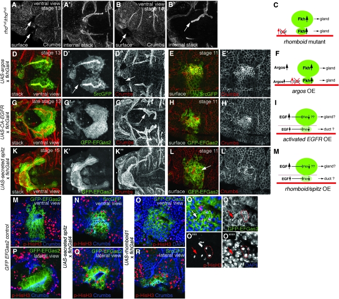
EGFR signaling is necessary but not sufficient to induce salivary duct fate, and overactivation leads to ectopic cell divisions. Analysis of components of the EGFR signaling pathway in the glands. It has been reported previously that salivary glands in rhomboid/spitz-group mutant embryos do not specify any ductal portion of the glands (Kuo et al. 1996). (A–B′) This phenotype was confirmed in a rhoPΔ5 embryos, a null allele of rho (Freeman et al. 1992). (A and A′) At stage 13, most of the secretory cells of the gland have invaginated, leaving two large holes visible at the surface of the embryo (arrows in A). (A) The surface of the embryo. (A′) An internal confocal stack to reveal the shape and location of the invaginated glands. (B and B′) At stage 14, the glands have fully invaginated and detached from the surface of the embryo, leaving no ductal connection to the outside and a large hole on the surface. (B) The surface of the embryo. (B′) An internal confocal stack to show the blunt-ended gland. The arrow in B′ points to the blunt end, and the arrow in B points to the hole. (A–B′) Labeling for Crumbs. (C) An amended gland-fate specification scheme as introduced in Figure 4 to illustrate the altered signaling in rho mutant embryos, where absence of EGFR signaling induces the entire salivary gland primordium to adopt secretory fate. (D–F) Overexpression of argos, an extracellular inhibitor of EGFR signaling (Schweitzer et al. 1995), using fkhGal4 caused a phenotype similar to that seen in rho mutants: (D–D″) At stage 13, the invaginated secretory portion of the glands detaches from the surface of the embryo (arrow in D′), and no duct is formed. (E and E′) The salivary gland primordium at stage 11 appears normal. (F) Scheme showing that downregulation of EGFR signaling leads to conversion of presumptive duct cells into secretory cells. (G–I) Overexpression of an activated form of the EGF receptor (CA-EGFR) using fkhGal4 leads to glands with highly disorganized and aberrant lumen from stage 12 onward. (G–G″) A confocal stack of stage 13 embryo; the arrow in G points to the lumen marked by Crumbs. (H and H′) Cell shapes marked by Crumbs in the salivary gland primordium at stage 11 appear normal. (I) Scheme showing that elevated EGFR signaling throughout the primordium does not induce duct fate in all cells. (K–I) Overexpression of a secreted and active form of the ligand Spitz using fkhGal4 leads to glands that very much resemble those seen upon overexpression of rho (compare with Figure 4). (K–K″) A confocal stack of a stage 15 embryo. No ductal structures are formed, and the shape of the secretory cells and the lumen is aberrant. (L and L′) The salivary gland primordium at stage 11 appears disrupted with irregular and too large apexes of the invaginating cells. The arrow in L points to a group of cells that show midbodies left by mitotic divisions marked by the GFP-EFGas2 microtubule marker. Ectopic mitoses can also be observed when CA-EGFR is expressed using fkhGal4. (M) Scheme showing that elevated EGFR signaling through overexpression of secreted Spitz throughout the primordium does not induce duct fate in all cells. The SrcGFP or GFP-EFGas2 markers are green in D, E, G, H, K, and L and shown as a single channel in D′, G′, and K′. Crumbs labeling is red in D, E, G, H, K, and L and shown as a single channel in D″, E′, G″, H′, K″, L′. (M–O″′) Analysis of ectopic cell divisions induced by activation of EGF signaling using phospho-histone H3 (p-HisH3) as a marker of mitosis. In the marker-expressing control, p-His3 labeling is restricted to the area outside the placode (M) and invaginated gland (P). When UAS-secreted spitz (N and Q) or UAS-rhomboid1 (O and R) are expressed in the glands, many mitotic cells can be found in the placode and invaginated gland. (M–R and O′) GFP markers, green; p-HisH3, red. (M–Q and R) Crumbs, blue. (O and O′) DAPI, blue. O′–O″″ show a higher magnification of the dividing cells in O. GFP-EFGas2 is shown as a single channel in O″, p-HisH3 as a single channel in O″′, and DAPI as a single channel in O″″. The dashed line in O′–O″″ highlights a cell in anaphase (note the spindle in O″) and the arrow in O′–O″″ points to a midbody in telophase, similar to the ones indicated in L.
Overexpression of rhomboid1 using EP(3)3704 in the glands led to the formation of glands that were positioned too anteriorly with no obvious distinction in shape between duct and secretory cells and no common duct-like structure formed at stage 15 (Figure 4, C–C″). An identical phenotype was observed when a rhomboid1 transgene under the control of the UAS promoter was expressed in the glands (Figure 4, D–D″). At earlier stages during the invagination process, when rhomboid1 was overexpressed in the gland primordium using fkhGal4 and either the EP(3)3704 or UAS-rhomboid1, aberrantly shaped lumena could be observed (Figure 4, E and E′), but most prominently a large bulge of fkh-expressing ectopic cells seemed to arise between the already invaginated secretory gland portions at the position where the individual ducts usually would form (Figure 4, F and F′; similar ectopic cells could also be observed in other experimental situations; see below). Although the analysis of rhomboid1 mutant embryos has shown that EGFR signaling is necessary to induce duct fate in the most ventral cells of the placode (and thus loss of EGFR signaling leads to loss of ducts; see Figure 5, A–B′), this could indicate that activation of EGFR signaling throughout the placode was not sufficient to induce duct fate in all cells. To test this hypothesis, we labeled embryos overexpressing rhomboid1 with markers for duct [Eye gone (Eyg); Jones et al. 1998] and secretory (dCreb-A; Abrams and Andrew 2005) gland fate and compared the expression to wild-type embryos. Both markers labeled the salivary placode (Figure 4, G and K) and the glands at stage 14 (Figure 4, H and L) and stage 15 (Figure 4, I and M) in a comparable pattern to wild-type placodes and glands. In addition, the bulge of potentially ectopic cells at the ventral surface that was observed at stages 13 and 14 (bracket in Figure 4L) strongly expressed the duct marker Eyg, indicating that cells fated to become duct cells have overproliferated. At stages 15 and 16, a large group of cells at the very anterior tip of the embryo that completely failed to invaginate expressed Eyg (Figure 4, M and M″). These data suggest that, at least when the EGFR pathway is ectopically activated throughout the whole placode in a time frame that mimicked the expression of fkh, this was not enough to convert secretory cells into duct cells. To test whether this failure in fate conversion could be due to the timing of the overexpression, we also expressed rhomboid1 under the control of armadilloGal4 (armGal4) throughout the whole epidermis of the embryo and with expression starting at much earlier stages (Figure 4, N–P″). The overactivation of EGFR signaling throughout the epidermis led to embryos with varying degrees of overall affected morphology (in many cases head involution failed, and general appearance of the epidermis was less organized compared to wild type, although epithelial integrity/polarity appeared unperturbed as judged by UAS-α-cateninGFP labeling that was also driven under the control of armGal4). In these embryos, dCreb-A and Eyg were expressed in the placode at stage 11, although the dCReb-A expression domain appeared to extend beyond the placode area into the more anterior hemi-segments (Figure 4, N and N′). Salivary gland invagination was strongly affected in that only very short glands invaginated into the embryo (see Figure 4, O and P, for stages 13 and 14, respectively). Nonetheless, these stubby glands expressed dCreb-A, the secretory fate marker, in the invaginated portion of the glands (Figure 4, O and P, arrows), and expressed Eyg in a few cells that had invaginated but were still close to the surface of the embryo (Figure 4P″). In addition, a large bulge of Eyg-expressing cells could be found at the surface of the embryo between the two invagination sides (Figure 4O, bracket), similar to what we observed when rhomboid1 was overexpressed under the control of fkhGal4.
We also tested this hypothesis further by overexpressing additional components of the EGF pathway in the salivary glands: a constitutively active version of the EGFR, UAS-CA-EGFR; a secreted version of the ligand Spitz, UAS-sspi; and the negative regulator Argos, UAS-argos. Overexpression of argos using fkhGal4 led to a high proportion of glands that lost a ductal connection to the embryo surface, similar to the rhomboid1 mutant embryos (Figure 5, A–D″), although less penetrant (which is probably due to timing and/or expression levels of the transgene). When a secreted version of the EGFR ligand Spitz was expressed using fkhGal4, the phenotypes observed appeared very similar to the ones seen in the rhomboid1 overexpressing embryos, namely ectopic cells and glands positioned too anteriorly without a discernible duct (Figure 5, K–L′). Overexpression of a constitutively active form of the EGFR, UAS-CA-EGFR, in the salivary glands led to invagination of cells with slightly aberrant shapes and to an invagination hole that was too large. This led to fully invaginated glands with a too large and aberrantly shaped lumen, although the individual and common ducts appeared normal (Figure 5, G–H′). The ectopic ventral cells observed upon expression of rhomboid1 or secreted spitz in the glands could be a result of overproliferation if EGFR signaling in the placodal cells works not only as a fate switch but also as a mitogenic signal. We thus analyzed the amount of cell division in the placode at stages 12 and 13 in embryos overexpressing rhomboid1 or secreted spitz under the control of fkhGal4 compared to wild-type placodes using an antibody against phosphorylated histone H3 (p-HisH3), a chromatin mark of mitotic cells (Wei et al. 1999). In control embryos, salivary placode cell nuclei at stages 12 and 13 did not contain nuclei showing p-HisH3 labeling (Figure 5, M and P), whereas many placode cells overexpressing secreted spitz (Figure 5, N and Q) or rhomboid1 (Figure 5, O and R) showed the p-HisH3 mark and were thus actively dividing. Also, labeling of microtubules with GFP-EFGas2 revealed mitotic figures (Figure 5, O′ and O″, dotted lines) and remnant spindle midbodies (Figure 5, O′ and O″, arrows).
The results presented above suggest the following: first, that EGFR signaling, although necessary for duct fate, is not sufficient to induce duct fate in all salivary placode cells, even though absence of EGFR signaling turns all cells into secretory cells. Second, an increase of EGFR signaling in the placode cells can induce excessive proliferation in a part of these cells, probably resulting in the mishapen glands observed upon rhomboid1 or spitz overexpression. Third, through modulation of fkh levels and downstream components within the placode, EGFR signaling might also impinge on the cell-shape changes that invaginating cells undergo.
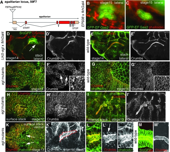
Egalitarian (Egl) and BicaudalD (BicD) are two proteins that act together with cytoplasmic Dynein in the localization of mRNAs in Drosophila embryos and the oocyte, with Egl interacting directly with Dynein light chain (Bullock and Ish-Horowicz 2001; Navarro et al. 2004). Overexpression of egl using EP(2)938 led to salivary glands that were C-shaped or shortened (Figure 6, B and C). Shortened glands could also be observed when egl was expressed in the glands using a UAS-egl construct (Figure 6, D and D′). GFP-positive cells that appeared to lose contact with the gland (arrow in Figure 6D) could be observed. Because both BicD and Egl have essential functions during oogenesis, an analysis of egl or BicD null embryos is not possible. We therefore analyzed embryos from mothers carrying two hypomorphic alleles of egl that were mated to heterozygous fathers (see materials and methods for the exact genotypes). Embryos with reduced Egl function often showed a disrupted epidermis, with large patches that appeared to completely lack apical Crumbs labeling (compare Figure 6, F and F′, to Figure 6, G and G′). This phenotype was variable, however, and an example of an embryo with a less disrupted epidermis is shown in Figure 6H. Also, during later stages of invagination at stage 13, the placode area was often disrupted and lacked apical Crumbs (Figure 6K). Salivary gland morphogenesis was disrupted in egl mutant embryos in that the cells of the placode often did not change their apices in a coordinated way (although crumbs still accumulated apically in the placode; see Figure 6H′), the invagination hole appeared too large and extended (Figure 6H), and the invaginated portion of the glands often had an irregular shape (Figure 6, I and K′). In the invaginated portion of a gland, Crumbs was not concentrated near the apical cell junctions as in the wild type, where this accumulation appears as a honeycomb lattice (compare Figure 6L′ to Figure 6M′). Instead, Crumbs was delocalized all over the apical surface and large accumulations could also be found intracellularly (arrow in Figure 6L′).
Figure 6.—
egalitarian overexpression reveals a potential role for egalitarian and BicD in salivary gland morphogenesis. (A) Scheme of the egalitarian (egl) locus indicating the gene structure and the position of the EP identified in the screen. (B and C) The two phenotypes observed in the screen upon overexpression of egl using EP(2)938: bent (B) and shortened (C) glands. The GFP-EFGas2 marker is in green, and phalloidin labeling is in red; both images are wide-field fluorescent. (D and D) Overexpression of a UAS-egl construct using fkhGal4 frequently led to short glands at stage 14, with some GFP-positive cells losing contact with the glands (arrow in D). (E and E′) A comparable wild-type embryo. Crumbs is in red in D and E and as a single channel in D′ and E′, SrcGFP is in green in D, and Shot is in green in E. (F–G′) Dorsolateral views of stage 14 embryos. (F and F′) egl mutant embryos often show a disrupted epidermis with mislocalized Crumbs labeling (arrows in F′ point to areas where Crumbs is completely absent), whereas in wild-type embryos Crumbs is localized apically and circumferentially in all epithelial cells (G and G′). (H and H′) egl mutant embryo showing a disorganized salivary gland placode, with a too-large and extended invagination hole. (I and I′) A stage 13 egl mutant embryo with a gland that appears too wide and short, showing mislocalized Crumbs labeling. (K and K′) Ventral view of a stage 13 egl mutant embryo. (K) A surface confocal stack, showing two disrupted areas in the epidermis where the glands have invaginated (arrows). (K′) An internal confocal stack of the same embryo, with a too-wide and aberrant gland (the red dashed line traces the outline of the gland). (L and L″) Higher magnification of the gland shown in K′ in a smaller confocal stack. Note the mislocalized Crumbs protein at the lateral sides of cells and internally (arrow in L′) that cannot be seen in wild-type glands (compare to M′). (H–L″) Crumbs labeling is red in H, I, K, and L and a single channel in H′, I′, K′, and L′; phalloidin labeling is green in H, I, K, and L and a single channel in L″. (M and M′) Magnification of a section through a stage 13 wild-type gland; SrcGFP is green in M, Crumbs is red in M and a single channel in M′.
What could be the mechanism leading to a loss of apical surface identity or constituents? Egl and BicD together with Dynein act as minus-end-directed microtubule motors, and as in most epithelial cells, the minus ends of microtubules are located near the apical surface in the salivary glands (Myat and Andrew 2002). hairy mRNA is one of the best-understood cargoes of Egl- and BicD-mediated transport (Bullock et al. 2003, 2006), and Hairy has been shown to be important for the regulation of apical membrane growth during salivary gland formation, in part through modulation of Crumbs (Myat and Andrew 2002). Thus, affecting hairy transcript localization through lowered levels of Egl and BicD could in turn affect the maintenance of apical membrane identity in the secretory cells. Alternatively, recent reports have shown that crumbs mRNA itself, and also the RNA of the Crumbs-associated protein Stardust (Std), are apically localized and this apical localization is important for function (Horne-Badovinac and Bilder 2008; Li et al. 2008). Thus, if crumbs mRNA localization were dependent upon Egl and BicD, then reduction of Egl and BicD would result in a loss of functional Crumbs at the apical surface, leading to a loss of epithelial characteristics. The apical localization at least of std mRNA appears developmentally regulated in the embryo (Horne-Badovinac and Bilder 2008). Thus, it is possible to envision that salivary gland cell apical maintenance is Egl and BicD dependent and especially sensitive to levels of Egl and thus to Crumbs in comparison to other epithelial tissues at the same stage. We are currently investigating this issue in more detail.
A gene endogenously expressed in glands identified through antisense expression:
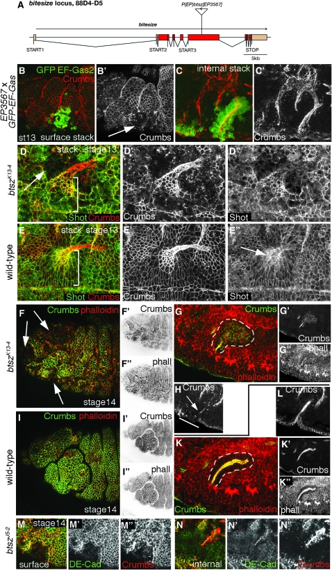
Bitesize (Btsz) is the sole Drosophila synaptotagmin-like protein. Its mRNA is expressed in the salivary glands and also in other epithelial tissues, with a strong apical enrichment (Serano and Rubin 2003). Btsz has recently been shown to control the organization of actin at adherens junctions in early embryos, although it might be dispensable in adult fly epithelia (Pilot et al. 2006). Recruitment of Btsz in early embryos to the apical junctional region is not dependent on E-Cadherin but on phosphatidylinositol-(4,5)-bisphosphate (PIP2) and Par-3/Bazooka, a protein of the Par-3/Par-6/aPKC apical complex (Pilot et al. 2006). Two mutant btsz alleles have been described: btszK13-4 that deletes a portion of the N terminus of some Btsz protein isoforms and btszJ5-2 that introduces a stop codon due to a frameshift in the N-terminal portion of btsz (Serano and Rubin 2003). Expression driven by fkhGal4 from the EP element identified in our screen, EP(3)3567, should lead to production of an antisense RNA in most of the btsz coding sequence and thus could downregulate endogenous btsz mRNA levels (see scheme in Figure 7A and supplemental Figure 2). In embryos where EP(3)3567 is driven by fkhGal4 at stage 13, when most secretory cells have invaginated from the placode, the epidermis in the region where the antisense RNA was expressed was disrupted and had lost apical Crumbs accumulation (Figure 7, B and B′). The glands themselves showed an irregular lumen (Figure 7, C and C′). btszK13-4 mutant embryos that manage to cellularize and complete gastrulation showed a somewhat disrupted epidermis, with loss of apical Crumbs accumulation in patches at later stages (see Figure 7D for a stage 13 and Figure 7F for a stage 14 embryo). Nonetheless, many salivary gland placode cells still showed enhanced Crumbs labeling, although the shapes of the apical circumferences of invaginating cells were irregular (compare Figure 7, D and D′, to wild type in Figure 7, E and E′). Also, the apical accumulation of the fly spectraplakin protein Shot could not be observed in btszK13-4 embryos in contrast to wild type (compare Figure 7D″ to Figure 7E″). At stage 14, the secretory portion of the glands in btszK13-4 embryos was often found to lose apical localization of Crumbs (and also to show reduced apical actin enrichment; Figure 7, G and G″). Similar to what we observed upon expression of EP(3)3567 with fkhGal4, the epidermis in the region where the placodal cells were located previously was disrupted and lost apical Crumbs and also DE-Cadherin labeling completely (compare Figure 7H to wild type in Figure 7L; Figure 7M). The disruption of the epidermis and the failure of the proper apical localization of Crumbs is to some extent reminiscent of the phenotypes observed in egl mutant embryos. As btsz mRNA is another RNA that is localized apically in various epithelial cells (and the localization signal has been identified; Serano and Rubin 2003), one could speculate that its localization could also be dependent on Egl and BicD, thus explaining some overlap in the phenotypes.
Figure 7.—
bitesize overexpression reveals a potential role for bitesize in salivary gland morphogenesis. (A) Scheme of the bitesize (btsz) locus indicating the gene structure and the position and orientation of the EP identified in the screen. (B–C′) Potential knockdown of Bitesize through overexpression of antisense RNA using EP(3)3567 leads to epithelial defects in the overexpressing cells (arrow in B′) and glands that invaginate with aberrant morphology. (B and B′) A surface confocal stack of a stage 13 embryo. (C and C′) The corresponding internal stack that reveals the glands. Note the absence of Crumbs labeling in the area that shows GFP-EFGas2 marker expression (arrow in B′). GFP-EFGas2 is green in B and C, Crumbs is red in B and C and a single channel in B′ and C′. (D–E′) Ventral views of btszK13-4 mutant vs. wild-type embryos at stage 13. The arrow in D points to the disrupted epidermis in the mutant embryo. Note the disorganization of the placode area compared to wild type (indicated by the brackets in D and E) and the failure to accumulate Shot apically (arrow in E″ indicates the accumulation in the wild type). Crumbs is red in D and E and a single channel in D′ and E′. Shot is green in D and E and a single channel in D″ and E″. (F–I″) Examples of lateral views of btszK13-4 mutant vs. wild-type embryos at stage 14. (F–F″) Highly disrupted and disorganized epidermis in the btszK13-4 mutant (arrows in F point to areas lacking apical circumferential Crumbs labeling; compare to the wild-type epidermis in I–I″). (G–G″) An internal stack of the same embryo as in F (the corresponding internal stack for the wild-type embryo in I is shown in K–K″). Note that the salivary gland of the btszK13-4 mutant embryo is losing apical Crumbs accumulation (G′) compared to the wild type (K′); the phalloidin labeling in G″ still shows cell outlines, but these also lack apical actin accumulation as seen in the wild type (K″). Crumbs labeling is green in F, G, I, and K and a single channel in F′, G′, I′, and K′, and phalloidin is red in F, G, I, and K and a single channel in F″, G″, I″, and K″. (H and L) Confocal stacks of the embryos in F and I at the level where the salivary duct reaches the epidermis labeled for Crumbs. (H) The btszK13-4 mutant. (L) The wild type. Note that the duct in H has lost apical Crumbs accumulation (the arrow points to the remnants of Crumbs labeling in the duct) and that the epidermis at the point from which the glands invaginated is disrupted and lacks apical Crumbs (indicated by the bar in H). (M–N″) The btszJ5-2 mutant at stage 14 also shows disrupted epidermis and loss of Crumbs (M″) and also DE-Cadherin (M′) in the area where the placode was previously located. Crumbs labeling in the invaginated gland is aberrant (N″) whereas DE-Cadherin still appears to be apical (N′). (M–M″) A projection of a 5-μm-thick confocal surface stack. (N–N″) The projection of a 20-μm-thick internal stack covering the whole gland. DE-Cadherin is green in M and N and a single channel in M′ and N′, and Crumbs is red in M and N and a single channel in M″ and N″.
A gene not endogenously expressed in the glands identified through overexpression:
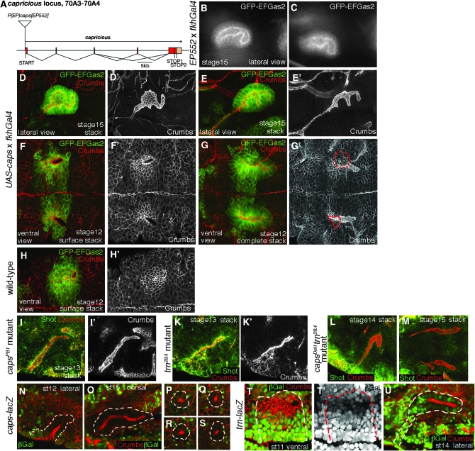
Capricious (Caps) belongs to the class of leucine-reach-repeat (LRR) transmembrane proteins, together with its close paralogue Tartan (Trn). Both proteins have been implicated in the formation of compartments of cells with different affinities in the wing disc (Milan et al. 2001), modulation of epithelial integrity within the wing disc (Mao et al. 2008), correct targeting of a subset of photoreceptor axons to the correct layer within the optic lobe (Shinza-Kameda et al. 2006), and the joining of tracheal branches over segment boundaries (Krause et al. 2006). One study also showed that in tissue culture Capricious and Tartan are able to mediate homophilic cell adhesion, a molecular function that could explain their roles discussed above (Shinza-Kameda et al. 2006). Overexpression of capricious using EP(3)552 led to glands with an enlarged lumen and very aberrant shapes at stage 15 (Figure 8, B and C). The same phenotype was observed when a transgene of capricious was expressed under UAS control (Figure 8, D–E′). During early stages of invagination, the invagination hole appeared enlarged compared to wild type and extended along the anterior–posterior axis (compare Figure 8, F–G′, to Figure 8, H and H′), suggesting problems in the shape of invagination cells and the order of invagination. This disorganization at later stages could lead to the aberrant shapes of the secretory part of the glands observed. We then analyzed capricious and tartan single and capricious and tartan double-mutant embryos (Mao et al. 2008), as some previous studies have indicated redundancy between both molecules in some tissues (Mao et al. 2008). In all mutant situations, invaginating glands often showed irregular lumens (Figure 8, I–M), indicating that both proteins might work together during salivary gland invagination. We next analyzed if and where capricious and tartan are expressed during salivary gland morphogenesis using P-element insertions in each gene that carry a lacZ gene leading to β-galactosidase expression under the endogenous expression control of each of the genes. Capricious was expressed in the embryo in the region of the salivary glands from stages 12–15, but appeared to be mostly excluded from the salivary glands themselves, although it was always expressed in cells in close contact with the glands (Figure 8, N–S). In contrast, tartan was expressed strongly in the salivary glands from placode stage onward (Figure 8, T and U). Similar expression patterns could also be observed in in situ hybridization for capricious and tartan mRNAs (see supplemental Figures 3 and 4). Thus, in analogy to the situation in the developing trachae where both proteins are expressed in complementary tissues to allow for proper dorsal branch fusion (Krause et al. 2006), the reciprocal expression of capricious and tartan in and around the salivary glands could play a part in the correct invagination and later positioning of the glands with respect to the surrounding tissues.
Figure 8.—
capricious overexpression reveals a potential role for capricious and tartan in salivary gland morphogenesis. (A) Scheme of the capricious (caps) locus indicating the gene structure and the position of the EP identified in the screen. (B and C) Live images of the GFP-EFGas2 marker of the caps overexpression phenotype using EP(3)552 observed in stage 15 embryos in the screen. (D–E′) Confocal stacks of two examples of aberrantly shaped lumen of salivary glands at stage 15 upon overexpression of a UAS-caps construct using fkhGal4. The lumen is highlighted by Crumbs labeling in D′ and E′ and very much resembles the defects observed in the screen. (F–H′) Invaginating glands at stage 12. (F and F′) A surface stack of the primordium upon UAS-caps overexpression. Note that the hole at the invagination point is too extended and not positioned completely within the primordium (as highlighted by the GFP marker) compared to the wild-type primordium in H and H′. (G and G′) A complete stack of the glands at stage 12 upon UAS-caps overexpression. Note that the size of the invagination hole (marked by the red dotted lines in G′) is again too large and irregular compared to wild type, and the invaginated portion of the glands shows a too-wide and irregular lumen. The GFP-EFGas2 marker is green and Crumbs labeling is red in D, E, F, G, and H and Crumbs is shown as a single channel in D′, E′, F′, and G′. (I–M) capsPB1 single, trn28.4 single, and capsDel1 trn28.4 double mutants (all are null mutations; Mao et al. 2008) often show defects in salivary gland morphology, i.e., irregular lumen at different stages of invagination. Staining for the fly spectraplakin Shot is green and Crumbs is red in I, K, L, and M, and Crumbs is shown as a single channel in I′ and K′. (N–S) A lacZ-containing P-element insertion into the caps locus reveals that caps is not expressed in most cells of the salivary glands. The outline of the glands is marked by a white dashed line, β-gal labeling is in green and Crumbs in red. N and O show β-galactosidase (β-gal) labeling at stage 12 and stage 15, respectively. (P–S) Cross sections of a gland at stage 14. Note that the glands are surrounded by cells expressing caps. (T–U) A lacZ-containing P-element insertion in the trn locus reveals that trn is expressed in salivary gland cells at all stages. (T and T′) Most cells of the salivary gland placode at stage 11 express trn at varying levels (border of the placode is marked by dashed lines). (U) At stage 14, trn is still expressed strongly in all salivary gland cells, including the duct. The outline of the gland is indicated by a dashed line. β-gal labeling is in green and Crumbs in red in T and U; β-gal is shown as a single channol in T′.
DISCUSSION
Here we show that a gain-of-function screen looking for factors affecting a process of epithelial morphogenesis, that is, the formation of the salivary glands in the Drosophila embryos, was efficient in identifying a range of known and new players. The screen was designed to identify genes that are endogenously expressed in the glands and where overexpression or antisense expression by an EP element interfered with endogenous function. The screen could also identify genes not endogenously expressed in the glands but with an apparent overexpression phenotype that uncovered a potential function for this gene in cell shape or cytoskeletal regulation elsewhere. Genes falling into all of these classes have been identified in the screen. We show that factors with a variety of proposed functions can affect salivary gland invagination—from cytoskeletal components via signaling factors (some of which impinge on the cytoskeleton themselves) to microRNAs and novel uncharacterized proteins. Most of the factors identified in this screen should affect salivary gland morphogenesis in a gland-autonomous fashion due to the restriction of overexpression to the salivary glands only, the only exception being the overexpression of secreted signaling factors.
We could observe a variety of phenotypes from aberrantly shaped glands to irregular lumena and wrongly positioned glands. In half of the cases, upon overexpression of a gene (or, in a few cases, potentially an antisense RNA) several different phenotypes could be observed, as opposed to a single dominant phenotype that was found in the other half. At the level of detail at which we analyzed the phenotypes (GFP markers of cell shape or microtubules plus phalloidin labeling of actin), these phenotypes seemed to fall into a limited number of classes, suggesting that several different perturbances of the system might lead to similar phenotypes. We also found no case that consistently led to a complete failure in salivary gland invagination. This is not completely surprising, as dominant effects or knockdowns that we would expect to see in our screen might not perturb the system enough to lead to a complete failure in all aspects of the cell-shape changes required for the invagination. Also, when analyzing various mutants in the initial follow-up of a subset of the hits identified, even in situations where the embryonic epidermis seemed to be very disrupted and cell shapes of invaginating cells were highly irregular, as, for example, in btsz or egl mutants, the glands nonetheless managed to invaginate. These observations suggest that there is a strong drive for the invagination of the cells of the salivary gland primordium, with many different factors contributing at the effector level. Elimination or perturbance of only one of these factors will not prevent invagination completely, but will rather lead to a slightly disordered invagination process that in the end might result in the aberrant shapes and phenotypes that we observed. This situation appears similar to the process of mesoderm invagination during gastrulation in the Drosophila embryo. Many factors contribute to this process, but loss of none—apart from the most upstream transcription factor initiating the whole mesoderm invagination program (twist)—will abolish invagination completely. In all other downstream mutants analyzed, the mesoderm will nonetheless manage to invaginate, albeit in an uncoordinated and/or delayed fashion (Leptin 2005). Similarly, during salivary gland invagiantion, fkh appears to be the most upstream transcription factor initiating the invagination program for both the secretory and the ductal part of the glands. In the absence of Fkh, invagination fails completely (Myat and Andrew 2000a). Several direct targets of Fkh have been identified, including the transcription factors senseless (Chandrasekaran and Beckendorf 2003) and sage (Abrams et al. 2006); PH4αSG2, a prolyl-4-hydroxylase (Abrams et al. 2006); and crebA, which in turns control the expression of secretory genes in the glands (Abrams and Andrew 2005).
Only one recent study has so far addressed the direct targets of fkh in a genomewide manner (Liu and Lehmann 2008). In this study, whole-genome expression levels in control pupae and pupae with forced expression of fkh were compared . At the beginning of pupariation, fkh controls both the expression of the salivary gland secretion proteins (Roth et al. 1999) and the cell death that occurs during pupariation in this tissue (Myat and Andrew 2000a). Downstream targets of fkh identified in this study included cell death genes and genes involved in autophagy, in phospholipid metabolism, in glucose and fatty acid metabolism, in hormone-dependent signaling pathways, and others. These other factors regulated by fkh included several proteins that we also identified in our screen: capricious, bitesize, gliotactin, ptpmeg, rhomboid1, and spitz. This overlap of fkh-dependent factors and genes identified by us, which are also potentially fkh dependent at different stages of development, i.e., at embryo and pupa stages, could suggest that these overlapping factors are regulated by fkh independently of stage-specific cofactors.
The set of genes identified in our screen encodes proteins with a wide range of potential functions: cytoskeleton or cytoskeleton associated, signaling, nuclear or transcription factor, protein synthesis and degradation, membrane traffic, cell surface and extracellular, enzymes, mitosis/meiosis germline, and uncharacterized genes and microRNAs. Nonetheless, many of these have been implicated in some aspect of epithelial morphogenetic function, some even within the salivary glands themselves. Thus, we are confident that many of the factors identified in this gain-of-function screen will turn out to have a function in epithelial morphogenesis. The initial folllow-up of the subset of genes described in detail above also confirms that gain-of-function phenotypes can point to new factors involved in a process and can also reveal new aspects of a function of a protein that were not previously appreciated, as in the case of rhomboid1.
The overexpression phenotype of rhomboid1 in the salivary glands showed an intriguing phenotype: ectopic EGFR signaling throughout the part of the primordium that will constitute the secretory part of the gland does not simply induce these cells to switch to ductal fate. Instead, it suggests either that other permissive factors that are absent from the secretory primordium are expressed in the duct primordium independently of EGFR signaling or that further inhibitory factors that prevent ductal fate are at work in the secretory primordium in addition to EGF-induced fkh. The timing of the overexpression of rhomboid1 in the screen, using part of the fkh promoter in the fkhGal4 line to drive the expression of the EP elements, did not seem to be crucial to the fate decision, as expression of rhomboid1 throughout the whole epidermis using an earlier Gal4-driver (armGal4) still led to invagination of glands with identifiable secretory and ductal cells (Figure 4, N–P). Thus it does not appear that the cells in the placode are responsive to the EGFR signal in terms of fate assignment in only a narrow window of time, but supports the notion that other factors are involved. It will be interesting to determine in the future what these factors are.
The analysis of caps and tartan mutant phenotypes suggests a role for these genes in salivary gland morphogenesis. The molecular function of both Caps and Trn proteins is still unclear. They appear important in mediating interaction and distinctiveness between groups of cells: neurons finding appropriate targets in the brain (Shishido et al. 1998; Shinza-Kameda et al. 2006), separation of ventral and dorsal compartment cells in the wing disc (Milan et al. 2001), and tracheal morphogenesis across segment boundaries (Krause et al. 2006). It has been suggested that Caps and Trn act as homophilic or heterophilic adhesion receptors or serve another unidentified function during adhesion. Our results indicate that salivary gland morphogenesis might be a useful system for addressing their molecular function in more detail. It is also interesting to note that not only Caps and Trn, but also Slit and the protein encoded by CG14351, which were also both identified as hits in the screen, belong to the family of LRR proteins, suggesting a general role for this class of surface receptors in salivary gland morphogenesis.
Another gene with a highly penetrant phenotype (severely shortened glands) identified in the screen is TNF-receptor-associated factor-4 (traf-4, previously annotated as traf-1 in FlyBase). Traf-4 has previously been shown to induce apoptosis via activation of JNK-kinase when overexpressed in other tissues (Kuranaga et al. 2002). Traf-4 has also been linked to the Ste20 kinase Mishapen (another gene identified in our screen), which in turn has been shown to be important for coordinated cell-shape changes occurring, for example, during dorsal closure in the fly embryo and during epiboly in the zebrafish embryo (Koppen et al. 2006). It also appears to have a role in mesoderm invagination (M. Leptin, personal communication). Overexpression of traf-4 using EP(2)578 led to a severe reduction in the number of cells in the salivary glands at embryonic stage 15 [when counting cell numbers in fluorescence images taken through the middle of wild-type glands, these had 39.9 ± 3.9 cells (n = 40) around the perimeter of the gland, whereas traf-4 overexpressing glands had 18.7 ± 5.0 cells (n = 45) around the perimeter]. We have not directly tested whether the missing cells died through induction of apoptosis, but would expect this to be the case in agreement with earlier studies. To address whether traf-4 has a function linked to mishapen and cell-shape changes in the glands, we analyzed fly embryos lacking Traf-4 (this mutant was a kind gift of Maria Leptin) for any problems in the early cell-shape changes occurring during salivary gland invagination, but could not find any strong defects (data not shown). Thus, despite many similarities between the epithelial morphogenetic processes of mesoderm invagination (which also starts with the invagination of an epithelial sheet) and salivary gland invagination, downstream effectors vary between the two systems.
Another interesting group of hits identified in the screen are the EP elements potentially driving overexpression of the mir-310 microRNA cluster. This cluster contains the microRNA genes mir-310, mir-311, mir-312, and mir-313. Overexpression of the cluster from three different EP elements located just upstream of the cluster, EP(2)2536, EP(2)2586, and EP(2)2587, led in each case to glands with widened and irregular lumena, although with varying penetrance (data not shown). Antisense-mediated depletion of each microRNA from this cluster has previously been shown to perturb dorsal closure and head involution in the embryo, indicating that the inhibition of downstream targets of this cluster might be important for various epithelial morphogenetic events (Leaman et al. 2005). Thus, the microRNAs in this cluster could be important in regulating targets that require downregulation to facilitate invagination during salivary gland morphogenesis. Two other microRNAs have been shown to be expressed in the salivary gland in the embryo—mir-8 and mir-375—with mir-8 showing a dynamic expression pattern (Aboobaker et al. 2005); expression patterns for the mir-310 cluster have not been analyzed yet. These data together with our screen results suggest that microRNA-dependent control of gene expression might be an important factor in salivary gland morphogenesis.
In summary, the gain-of-function screen for factors affecting cell shape during salivary gland morphogenesis in the Drosophila embryo presented here was successful in identifying a range of candidates. These candidates represent, on the one hand, genes that are endogenously expressed in the glands and thus are likely to serve a role during salivary gland morphogenesis/tubulogenesis. On the other hand, we identified genes that are not endogenously expressed in the glands but nonetheless interfered with their invagination, potentially through effects on cell shape or the cytoskeleton. These genes might therefore also be important for the regulation of cell shape in other tissues. It will be interesting to determine which of the candidate genes of the first class serve a function only in the salivary glands and which are required for tubulogenesis events in general and in other species. For the second group of candidates, an analysis of their role during cell-shape changes in other morphogenetic events will be key to understanding how they affected salivary gland morphogenesis in our screen. Because the screen identified several uncharacterized genes whose expression gave strong phenotypes in the glands and that have close orthologs in mammals, the salivary glands appear to be a good model system for analyzing the function of such genes. Finally, our follow-up investigation of a selected set of candidate genes through loss-of-function mutants demonstrates that the combination of functional screening and phenotypic loss-of-function analysis provides a useful approach to identifying downstream effectors in a morphogenetic process.
Acknowledgments
The authors thank Nick Brown, Matthew Freeman, Simon Bullock, Deborah Andrew, Maria Leptin, and the Bloomington and Szeged Stock Centers for fly stocks; Matthew Freeman, Simon Bullock, Sarah Bray, Deborah Andrew, and the Developmental Studies Hybridoma Bank at the University of Iowa for antibodies; Maria Leptin for communication of results prior to publication; Nick Brown for use of his confocal microscope; and Sean Munro and Nick Brown for helpful comments on the manuscript. This work was supported by the Biotechnology and Biological Sciences Research Council (grant no. BB/B501798/1) and the Royal Society.
References
- Aboobaker, A. A., P. Tomancak, N. Patel, G. M. Rubin and E. C. Lai, 2005. Drosophila microRNAs exhibit diverse spatial expression patterns during embryonic development. Proc. Natl. Acad. Sci. USA 102 18017–18022. [DOI] [PMC free article] [PubMed] [Google Scholar]
- Abrams, E. W., and D. J. Andrew, 2005. CrebA regulates secretory activity in the Drosophila salivary gland and epidermis. Development 132 2743–2758. [DOI] [PubMed] [Google Scholar]
- Abrams, E. W., W. K. Mihoulides and D. J. Andrew, 2006. Fork head and Sage maintain a uniform and patent salivary gland lumen through regulation of two downstream target genes, PH4alphaSG1 and PH4alphaSG2. Development 133 3517–3527. [DOI] [PubMed] [Google Scholar]
- Andrew, D. J., A. Baig, P. Bhanot, S. M. Smolik and K. D. Henderson, 1997. The Drosophila dCREB-A gene is required for dorsal/ventral patterning of the larval cuticle. Development 124 181–193. [DOI] [PubMed] [Google Scholar]
- Barros, T. P., K. Kinoshita, A. A. Hyman and J. W. Raff, 2005. Aurora A activates D-TACC-Msps complexes exclusively at centrosomes to stabilize centrosomal microtubules. J. Cell Biol. 170 1039–1046. [DOI] [PMC free article] [PubMed] [Google Scholar]
- Bate, M., and A. Martinez Arias, 1993. The Development of Drosophila melanogaster, Vol. II. Cold Spring Harbor Laboratory Press, Cold Spring Harbor, NY.
- Bejarano, F., C. M. Luque, H. Herranz, G. Sorrosal, N. Rafel et al., 2008. A gain-of-function suppressor screen for genes involved in dorsal-ventral boundary formation in the Drosophila wing. Genetics 178 307–323. [DOI] [PMC free article] [PubMed] [Google Scholar]
- Benlali, A., I. Draskovic, D. J. Hazelett and J. E. Treisman, 2000. act up controls actin polymerization to alter cell shape and restrict Hedgehog signaling in the Drosophila eye disc. Cell 101 271–281. [DOI] [PubMed] [Google Scholar]
- Bornemann, D. J., J. E. Duncan, W. Staatz, S. Selleck and R. Warrior, 2004. Abrogation of heparan sulfate synthesis in Drosophila disrupts the Wingless, Hedgehog and Decapentaplegic signaling pathways. Development 131 1927–1938. [DOI] [PubMed] [Google Scholar]
- Bradley, P. L., M. M. Myat, C. A. Comeaux and D. J. Andrew, 2003. Posterior migration of the salivary gland requires an intact visceral mesoderm and integrin function. Dev. Biol. 257 249–262. [DOI] [PubMed] [Google Scholar]
- Bray, S., H. Musisi and M. Bienz, 2005. Bre1 is required for Notch signaling and histone modification. Dev. Cell 8 279–286. [DOI] [PubMed] [Google Scholar]
- Bullock, S. L., and D. Ish-Horowicz, 2001. Conserved signals and machinery for RNA transport in Drosophila oogenesis and embryogenesis. Nature 414 611–616. [DOI] [PubMed] [Google Scholar]
- Bullock, S. L., D. Zicha and D. Ish-Horowicz, 2003. The Drosophila hairy RNA localization signal modulates the kinetics of cytoplasmic mRNA transport. EMBO J. 22 2484–2494. [DOI] [PMC free article] [PubMed] [Google Scholar]
- Bullock, S. L., A. Nicol, S. P. Gross and D. Zicha, 2006. Guidance of bidirectional motor complexes by mRNA cargoes through control of dynein number and activity. Curr. Biol. 16 1447–1452. [DOI] [PubMed] [Google Scholar]
- Campos-Ortega, J. A., and V. Hartenstein, 1985. The Embryonic Development of Drosophila melanogaster. Springer-Verlag, Berlin.
- Cha, G. H., K. S. Cho, J. H. Lee, M. Kim, E. Kim et al., 2003. Discrete functions of TRAF1 and TRAF2 in Drosophila melanogaster mediated by c-Jun N-terminal kinase and NF-kappaB-dependent signaling pathways. Mol. Cell. Biol. 23 7982–7991. [DOI] [PMC free article] [PubMed] [Google Scholar]
- Chandrasekaran, V., and S. K. Beckendorf, 2003. senseless is necessary for the survival of embryonic salivary glands in Drosophila. Development 130 4719–4728. [DOI] [PubMed] [Google Scholar]
- Chandrasekaran, V., and S. K. Beckendorf, 2005. Tec29 controls actin remodeling and endoreplication during invagination of the Drosophila embryonic salivary glands. Development 132 3515–3524. [DOI] [PubMed] [Google Scholar]
- Colas, J. F., and G. C. Schoenwolf, 2001. Towards a cellular and molecular understanding of neurulation. Dev. Dyn. 221 117–145. [DOI] [PubMed] [Google Scholar]
- Daubresse, G., R. Deuring, L. Moore, O. Papoulas, I. Zakrajsek et al., 1999. The Drosophila kismet gene is related to chromatin-remodeling factors and is required for both segmentation and segment identity. Development 126 1175–1187. [DOI] [PubMed] [Google Scholar]
- Florence, B., A. Guichet, A. Ephrussi and A. Laughon, 1997. Ftz-F1 is a cofactor in Ftz activation of the Drosophila engrailed gene. Development 124 839–847. [DOI] [PubMed] [Google Scholar]
- Freeman, M., B. E. Kimmel and G. M. Rubin, 1992. Identifying targets of the rough homeobox gene of Drosophila: evidence that rhomboid functions in eye development. Development 116 335–346. [DOI] [PubMed] [Google Scholar]
- Haberman, A. S., D. D. Isaac and D. J. Andrew, 2003. Specification of cell fates within the salivary gland primordium. Dev. Biol. 258 443–453. [DOI] [PubMed] [Google Scholar]
- Harris, K. E., and S. K. Beckendorf, 2007. Different Wnt signals act through the Frizzled and RYK receptors during Drosophila salivary gland migration. Development 134 2017–2025. [DOI] [PubMed] [Google Scholar]
- Harris, K. E., N. Schnittke and S. K. Beckendorf, 2007. Two ligands signal through the Drosophila PDGF/VEGF receptor to ensure proper salivary gland positioning. Mech. Dev. 124 441–448. [DOI] [PMC free article] [PubMed] [Google Scholar]
- Henderson, K. D., and D. J. Andrew, 2000. Regulation and function of Scr, exd, and hth in the Drosophila salivary gland. Dev. Biol. 217 362–374. [DOI] [PubMed] [Google Scholar]
- Henderson, K. D., D. D. Isaac and D. J. Andrew, 1999. Cell fate specification in the Drosophila salivary gland: the integration of homeotic gene function with the DPP signaling cascade. Dev. Biol. 205 10–21. [DOI] [PubMed] [Google Scholar]
- Hogan, B. L., and P. A. Kolodziej, 2002. Organogenesis: molecular mechanisms of tubulogenesis. Nat. Rev. Genet. 3 513–523. [DOI] [PubMed] [Google Scholar]
- Horne-Badovinac, S., and D. Bilder, 2008. Dynein regulates epithelial polarity and the apical localization of stardust A mRNA. PLoS Genet. 4 e8. [DOI] [PMC free article] [PubMed] [Google Scholar]
- Houalla, T., D. Hien Vuong, W. Ruan, B. Suter and Y. Rao, 2005. The Ste20-like kinase misshapen functions together with Bicaudal-D and dynein in driving nuclear migration in the developing Drosophila eye. Mech. Dev. 122 97–108. [DOI] [PubMed] [Google Scholar]
- Jones, N. A., Y. M. Kuo, Y. H. Sun and S. K. Beckendorf, 1998. The Drosophila Pax gene eye gone is required for embryonic salivary duct development. Development 125 4163–4174. [DOI] [PubMed] [Google Scholar]
- Kaltschmidt, J. A., C. M. Davidson, N. H. Brown and A. H. Brand, 2000. Rotation and asymmetry of the mitotic spindle direct asymmetric cell division in the developing central nervous system. Nat. Cell Biol. 2 7–12. [DOI] [PubMed] [Google Scholar]
- Kolesnikov, T., and S. K. Beckendorf, 2005. NETRIN and SLIT guide salivary gland migration. Dev. Biol. 284 102–111. [DOI] [PubMed] [Google Scholar]
- Koppen, M., B. G. Fernandez, L. Carvalho, A. Jacinto and C. P. Heisenberg, 2006. Coordinated cell-shape changes control epithelial movement in zebrafish and Drosophila. Development 133 2671–2681. [DOI] [PubMed] [Google Scholar]
- Krause, C., C. Wolf, J. Hemphala, C. Samakovlis and R. Schuh, 2006. Distinct functions of the leucine-rich repeat transmembrane proteins capricious and tartan in the Drosophila tracheal morphogenesis. Dev. Biol. 296 253–264. [DOI] [PubMed] [Google Scholar]
- Kuo, Y. M., N. Jones, B. Zhou, S. Panzer, V. Larson et al., 1996. Salivary duct determination in Drosophila: roles of the EGF receptor signalling pathway and the transcription factors fork head and trachealess. Development 122 1909–1917. [DOI] [PubMed] [Google Scholar]
- Kuranaga, E., H. Kanuka, T. Igaki, K. Sawamoto, H. Ichijo et al., 2002. Reaper-mediated inhibition of DIAP1-induced DTRAF1 degradation results in activation of JNK in Drosophila. Nat. Cell Biol. 4 705–710. [DOI] [PubMed] [Google Scholar]
- Kusano, A., C. Staber and B. Ganetzky, 2001. Nuclear mislocalization of enzymatically active RanGAP causes segregation distortion in Drosophila. Dev. Cell 1 351–361. [DOI] [PubMed] [Google Scholar]
- Leaman, D., P. Y. Chen, J. Fak, A. Yalcin, M. Pearce et al., 2005. Antisense-mediated depletion reveals essential and specific functions of microRNAs in Drosophila development. Cell 121 1097–1108. [DOI] [PubMed] [Google Scholar]
- Leptin, M., 2005. Gastrulation movements: the logic and the nuts and bolts. Dev. Cell 8 305–320. [DOI] [PubMed] [Google Scholar]
- Li, Z., L. Wang, T. S. Hays and Y. Cai, 2008. Dynein-mediated apical localization of crumbs transcripts is required for Crumbs activity in epithelial polarity. J. Cell Biol. 180 31–38. [DOI] [PMC free article] [PubMed] [Google Scholar]
- Liu, Y., and M. Lehmann, 2008. Genes and biological processes controlled by the Drosophila FOXA orthologue Fork head. Insect Mol. Biol. 17 91–101. [DOI] [PubMed] [Google Scholar]
- Lubarsky, B., and M. A. Krasnow, 2003. Tube morphogenesis: making and shaping biological tubes. Cell 112 19–28. [DOI] [PubMed] [Google Scholar]
- Mao, Y., M. Kerr and M. Freeman, 2008. Modulation of Drosophila retinal epithelial integrity by the adhesion proteins capricious and tartan. PLoS ONE 3 e1827. [DOI] [PMC free article] [PubMed] [Google Scholar]
- Milan, M., U. Weihe, L. Perez and S. M. Cohen, 2001. The LRR proteins capricious and Tartan mediate cell interactions during DV boundary formation in the Drosophila wing. Cell 106 785–794. [DOI] [PubMed] [Google Scholar]
- Minakhina, S., R. Myers, M. Druzhinina and R. Steward, 2005. Crosstalk between the actin cytoskeleton and Ran-mediated nuclear transport. BMC Cell Biol. 6 32. [DOI] [PMC free article] [PubMed] [Google Scholar]
- Molnar, C., A. Lopez-Varea, R. Hernandez and J. F. de Celis, 2006. A gain-of-function screen identifying genes required for vein formation in the Drosophila melanogaster wing. Genetics 174 1635–1659. [DOI] [PMC free article] [PubMed] [Google Scholar]
- Myat, M. M., and D. J. Andrew, 2000. a Fork head prevents apoptosis and promotes cell shape change during formation of the Drosophila salivary glands. Development 127 4217–4226. [DOI] [PubMed] [Google Scholar]
- Myat, M. M., and D. J. Andrew, 2000. b Organ shape in the Drosophila salivary gland is controlled by regulated, sequential internalization of the primordia. Development 127 679–691. [DOI] [PubMed] [Google Scholar]
- Myat, M. M., and D. J. Andrew, 2002. Epithelial tube morphology is determined by the polarized growth and delivery of apical membrane. Cell 111 879–891. [DOI] [PubMed] [Google Scholar]
- Navarro, C., H. Puthalakath, J. M. Adams, A. Strasser and R. Lehmann, 2004. Egalitarian binds dynein light chain to establish oocyte polarity and maintain oocyte fate. Nat. Cell Biol. 6 427–435. [DOI] [PubMed] [Google Scholar]
- Panzer, S., D. Weigel and S. K. Beckendorf, 1992. Organogenesis in Drosophila melanogaster: embryonic salivary gland determination is controlled by homeotic and dorsoventral patterning genes. Development 114 49–57. [DOI] [PubMed] [Google Scholar]
- Paul, S. M., M. Ternet, P. M. Salvaterra and G. J. Beitel, 2003. The Na+/K+ ATPase is required for septate junction function and epithelial tube-size control in the Drosophila tracheal system. Development 130 4963–4974. [DOI] [PubMed] [Google Scholar]
- Pilot, F., J. M. Philippe, C. Lemmers and T. Lecuit, 2006. Spatial control of actin organization at adherens junctions by a synaptotagmin-like protein Btsz. Nature 442 580–584. [DOI] [PubMed] [Google Scholar]
- Pirraglia, C., R. Jattani and M. M. Myat, 2006. Rac function in epithelial tube morphogenesis. Dev. Biol. 290 435–446. [DOI] [PubMed] [Google Scholar]
- Rørth, P., K. Szabo, A. Bailey, T. Laverty, J. Rehm et al., 1998. Systematic gain-of-function genetics in Drosophila. Development 125 1049–1057. [DOI] [PubMed] [Google Scholar]
- Roth, G. E., S. Wattler, H. Bornschein, M. Lehmann and G. Korge, 1999. Structure and regulation of the salivary gland secretion protein gene Sgs-1 of Drosophila melanogaster. Genetics 153 753–762. [DOI] [PMC free article] [PubMed] [Google Scholar]
- Schenkel, H., S. Hanke, C. De Lorenzo, R. Schmitt and B. M. Mechler, 2002. P elements inserted in the vicinity of or within the Drosophila snRNP SmD3 gene nested in the first intron of the ornithine decarboxylase antizyme gene affect only the expression of SmD3. Genetics 161 763–772. [DOI] [PMC free article] [PubMed] [Google Scholar]
- Schweitzer, R., R. Howes, R. Smith, B. Z. Shilo and M. Freeman, 1995. Inhibition of Drosophila EGF receptor activation by the secreted protein Argos. Nature 376 699–702. [DOI] [PubMed] [Google Scholar]
- Serano, J., and G. M. Rubin, 2003. The Drosophila synaptotagmin-like protein bitesize is required for growth and has mRNA localization sequences within its open reading frame. Proc. Natl. Acad. Sci. USA 100 13368–13373. [DOI] [PMC free article] [PubMed] [Google Scholar]
- Shilo, B. Z., 2005. Regulating the dynamics of EGF receptor signaling in space and time. Development 132 4017–4027. [DOI] [PubMed] [Google Scholar]
- Shinza-Kameda, M., E. Takasu, K. Sakurai, S. Hayashi and A. Nose, 2006. Regulation of layer-specific targeting by reciprocal expression of a cell adhesion molecule, capricious. Neuron 49 205–213. [DOI] [PubMed] [Google Scholar]
- Shishido, E., M. Takeichi and A. Nose, 1998. Drosophila synapse formation: regulation by transmembrane protein with Leu-rich repeats, CAPRICIOUS. Science 280 2118–2121. [DOI] [PubMed] [Google Scholar]
- Steigemann, P., A. Molitor, S. Fellert, H. Jackle and G. Vorbruggen, 2004. Heparan sulfate proteoglycan syndecan promotes axonal and myotube guidance by slit/robo signaling. Curr. Biol. 14 225–230. [DOI] [PubMed] [Google Scholar]
- Strumpf, D., and T. Volk, 1998. Kakapo, a novel cytoskeletal-associated protein is essential for the restricted localization of the neuregulin-like factor, vein, at the muscle-tendon junction site. J. Cell Biol. 143 1259–1270. [DOI] [PMC free article] [PubMed] [Google Scholar]
- Subramanian, A., A. Prokop, M. Yamamoto, K. Sugimura, T. Uemura et al., 2003. Shortstop recruits EB1/APC1 and promotes microtubule assembly at the muscle-tendon junction. Curr. Biol. 13 1086–1095. [DOI] [PubMed] [Google Scholar]
- Svendsen, P. C., S. D. Marshall, M. Kyba and W. J. Brook, 2000. The combgap locus encodes a zinc-finger protein that regulates cubitus interruptus during limb development in Drosophila melanogaster. Development 127 4083–4093. [DOI] [PubMed] [Google Scholar]
- Szul, T., R. Grabski, S. Lyons, Y. Morohashi, S. Shestopal et al., 2007. Dissecting the role of the ARF guanine nucleotide exchange factor GBF1 in Golgi biogenesis and protein trafficking. J. Cell Sci. 120 3929–3940. [DOI] [PubMed] [Google Scholar]
- Tautz, D., and C. Pfeifle, 1989. A non-radioactive in situ hybridization method for the localization of specific RNAs in Drosophila embryos reveals translational control of the segmentation gene hunchback. Chromosoma 98 81–85. [DOI] [PubMed] [Google Scholar]
- The, I., Y. Bellaiche and N. Perrimon, 1999. Hedgehog movement is regulated through tout velu-dependent synthesis of a heparan sulfate proteoglycan. Mol. Cell 4 633–639. [DOI] [PubMed] [Google Scholar]
- Townsley, F. M., and M. Bienz, 2000. Actin-dependent membrane association of a Drosophila epithelial APC protein and its effect on junctional Armadillo. Curr. Biol. 10 1339–1348. [DOI] [PubMed] [Google Scholar]
- Verheyen, E. M., and L. Cooley, 1994. Profilin mutations disrupt multiple actin-dependent processes during Drosophila development. Development 120 717–728. [DOI] [PubMed] [Google Scholar]
- Vining, M. S., P. L. Bradley, C. A. Comeaux and D. J. Andrew, 2005. Organ positioning in Drosophila requires complex tissue-tissue interactions. Dev. Biol. 287 19–34. [DOI] [PubMed] [Google Scholar]
- Wei, Y., L. Yu, J. Bowen, M. A. Gorovsky and C. D. Allis, 1999. Phosphorylation of histone H3 is required for proper chromosome condensation and segregation. Cell 97 99–109. [DOI] [PubMed] [Google Scholar]
- Xu, H., J. A. Brill, J. Hsien, R. McBride, G. L. Boulianne et al., 2002. Syntaxin 5 is required for cytokinesis and spermatid differentiation in Drosophila. Dev. Biol. 251 294–306. [DOI] [PubMed] [Google Scholar]
- Xu, N., B. Keung and M. M. Myat, 2008. Rho GTPase controls invagination and cohesive migration of the Drosophila salivary gland through Crumbs and Rho-kinase. Dev. Biol. 321 88–100. [DOI] [PubMed] [Google Scholar]
- Yarmola, E. G., and M. R. Bubb, 2006. Profilin: emerging concepts and lingering misconceptions. Trends Biochem. Sci. 31 197–205. [DOI] [PubMed] [Google Scholar]
- Zhou, B., A. Bagri and S. K. Beckendorf, 2001. Salivary gland determination in Drosophila: a salivary-specific, fork head enhancer integrates spatial pattern and allows fork head autoregulation. Dev. Biol. 237 54–67. [DOI] [PubMed] [Google Scholar]
- Zhu, S., R. Perez, M. Pan and T. Lee, 2005. Requirement of Cul3 for axonal arborization and dendritic elaboration in Drosophila mushroom body neurons. J. Neurosci. 25 4189–4197. [DOI] [PMC free article] [PubMed] [Google Scholar]


