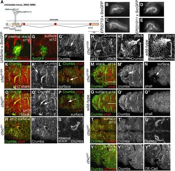
Figure 3.—
Chickadee (Profilin) is important for salivary gland invagination. chickadee encodes the Drosophila Profilin protein. (A) Scheme of the chic locus indicating the position and orientation of the two EP lines that showed phenotypes when driven in the salivary glands. B and C and D and E show phenotypes observed in the screen for EP713 and EP1011 respectively; wide-field fluorescent images of live embryos are shown. (F–G′) Overexpression of chickadee using a UAS-chickadee construct led to invagination problems and aberrantly shaped glands. (F) An internal confocal stack of a gland (14 μm thick), labeled with Crumbs to reveal the apical surface/lumen of the glands (red) and showing the SrcGFP marker in green. (G and G′) A surface stack of the same embryo (with Crumbs in red and SrcGFP in green in G and Crumbs as a single channel in G′; 3 μm thick). The arrow G′points to disrupted epidermis in the region of the placode from which the glands have started to invaginate. Note the absence of Crumbs from the apical surface of cells in this region. (H and H′) A low and high magnification view, respectively, of the slightly disorganized epidermis of a chic mutant embryo labeled for crumbs, with H′ showing the salivary gland placode and gland in a projection (18-μm thick stack). (I) A wild-type placode and gland (17-μm thick stack) at the same stage as in H′. Note that the highly organized arrangement of apical constriction of the placodal cells is less apparent in the chic mutant (bracket in H′ and in I). K and L and O and P show the aberrant glands and disrupted epidermis in two different chic alleles (chic01320 and chic221) at stage 12; labeling for Crumbs is in green (and also shown as a single channel in K′ and O′) and for phalloidin in red. Note the disruption and absence of apical Crumbs labeling in the region of the salivary gland placode (arrows in K′, L, O′, and P point to these areas). (K) A projection of a 35-μm thick stack. (L) A 5-μm-thick surface projection. (O) A projection of a 26-μm-thick stack. (P) A 3-μm-thick surface projection. (M and N) The disrupted epidermis in the region from which salivary gland cells invaginated in a stage 14 embryo. Crumbs is green in M and a single channel in M′, and phalloidin is red in M and a single channel in N. M is a projection of a 34-μm-thick stack, and N is a 5-μm-thick surface stack. For comparison, a stage 14 wild-type embryo is shown in Q–Q″. Crumbs is green in Q and a single channel in Q′, and phalloidin is red in Q and a single channel in Q″; Q is a 5-μm-thick surface stack. The arrows in M′ and N point to the disrupted region, the white lines in M and Q indicate the ventral midline (the view in M–N is slightly oblique). (R–V″) chic221 mutant embryos at stages 12–14. R and R′ show lateral views of a placode, whereas S shows an internal stack of the gland. T and T′ are ventral views of the two placodes, with U showing an internal stack of the glands. (V–V″) A surface stack of a mutant embryo (5 μm thick). Crumbs is green in R, T, and V and a single channel in R′, T′, and V′; phalloidin is red. Both S and U show Crumbs labeling to outline the lumen of the gland. DE-Cadherin (DE-Cad) labeling is in red in V and as a single channel in V″.

