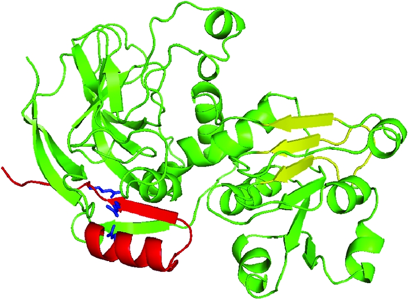
Figure 5.—

Ribbon diagram representation for the structure of SbCAD2. The significant arginine mutations found in SbCAD2_27b are indicated in blue. The dimer contact positions for βD–F are highlighted in yellow. The three secondary structural elements, β11, β12, and α6 are not maintained in the CAD model of bmr6-27 mutant due to the degenerative hydrophobic interactions caused by the arginine residues highlighted in blue.
