
Figure 6. Lack of primary cilia in Δ1-3 but not Δ2-5 homozygous mouse embryo fibroblasts.
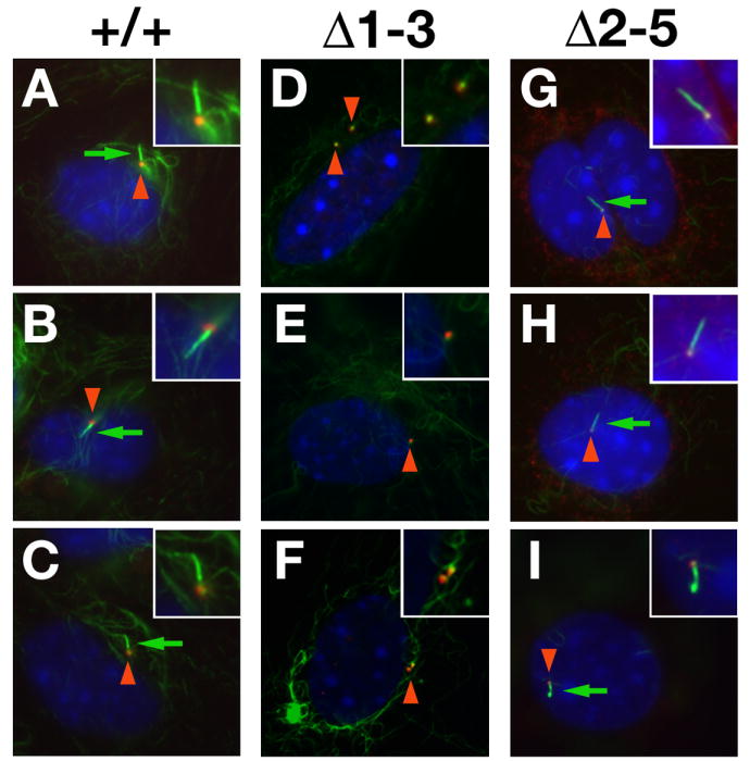
Wild type (A-C), Δ1-3 (D-F) and Δ2-5 homozygous (G-H) MEFs, serum starved for 48 hours, were immunostained with antibodies against acetylated tubulin (green) and γ-tubulin (red), whereas nuclei were counterstained with DAPI. Basal bodies (red arrowheads) were detected in both genotypes whereas primary cilia (green arrows) were only detected in wild type and Δ2-5 MEFs. Insets show enlargements of basal bodies and cilia.