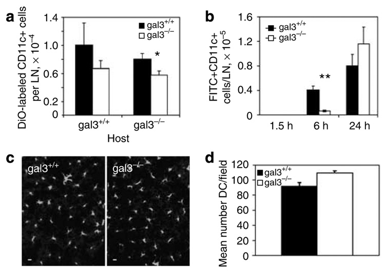
Figure 3. Impaired in vivo migration of bone marrow-derived immature dendritic cells from galectin-3-deficient mice.
(a) DiOC16-labeled BMiDCs (2 × 10⁶) from gal3−/− and gal3+/+ mice were injected into contralateral footpads of gal3+/+ and gal3−/− mice. Fluorescent CD11c+ DCs in inguinal lymph nodes were counted by flow cytometry 2 days later. Data are means ± SE from one of three experiments. *P<0.05. (b) Fluorescein isothiocyanate was applied to the lower abdomens of gal3−/− and gal3+/+ mice, and CD11c+ lymph node cells from digested inguinal nodes were analyzed by flow cytometry at the indicated periods. At time 0, cells were isolated immediately after fluorescein application. Data are means ± SE from a representative experiment of two. Four to six mice per genotype were used in each experiment. **P<0.01. (c) Major histocompatibility complex class II+ cells in epidermis from gal3−/− and gal3+/+ mouse pinnae were visualized by epifluorescence microscopy on an Olympus IX61 with a × 20 objective (NA 0.7). Bars represent a distance of 10 µm. Contrasts were adjusted identically to improve visibility. (d) Numbers of epidermal DCs from five contiguous micrograph fields (original magnification × 20) are shown for gal3−/− and gal3+/+ mice, means ± SE.

