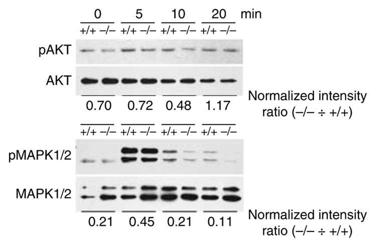
Figure 5. Immature gal3−/− DCs exhibit signaling defects in response to chemokine.
Gal3+/+ and gal3−/− BMiDCs were activated with 2.5 µm fMLP and equivalent cell numbers were harvested at the indicated periods. Cell lysates were processed for immunoblotting analysis to detect phosphorylated proteins and total protein. Numbers underneath each panel represent ratios of normalized phosphoprotein levels in gal3−/− cells divided by the corresponding levels in gal3+/+ cells.

