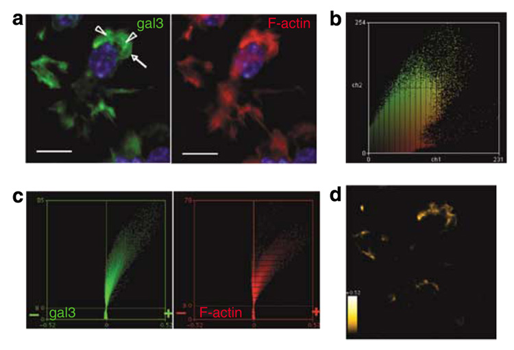
Figure 6. Galectin-3 is localized in membrane ruffles of immature bone marrow-derived dendritic cells.
BMiDC motility was induced by adhesion on fibronectin-coated coverslips and exposure to fMLP. (a) Detection of intracellular galectin-3 with antibody in the presence of Fc-Block, and F-actin with labeled phalloidin in the projection image. Fluorescence micrographs were acquired in permeabilized cells with a × 60 objective (NA 1.2) at intervals of 0.5 µm and deconvolved from optical planes of the entire cell. Images are contrast enhanced to improve visibility. Arrowheads and arrows indicate ruffles and lamellipodia, respectively. The bar represents a distance of 10 mm. (b) Diagonal distribution of galectin-3 (green) and F-actin (red) staining of all optical planes from unenhanced images of the field depicted in (a) indicates co-dependent pixel intensities. (c) Intensity correlation analyses of thresholded unenhanced images show pronounced positive skewing of PDM (product difference of the means, see Materials and Methods) values for galectin-3 and F-actin, indicative of significant co-localization. (d) Regions of the cell demonstrating galectin-3 and F-actin co-localization from positive PDM guidance in (c) are shown. Heat scale, PDM values 0 to ±0.52. Image is contrast enhanced to improve visibility. Results are representative of two experiments.

