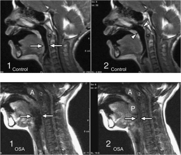
Figure 1.
Top: Three-year-old normal control male subject. Sagittal images from various points in the respiratory cycle show no significant change in airway diameter at the level of the hypopharynx (arrows in image 1) or nasopharynx (arrowheads in image 2). Bottom: Eleven-year-old male patient with obstructive sleep apnea (OSA). Sagittal images from various points of the respiratory cycle demonstrate airway collapse at the level of the hypopharynx (arrows in images 1 and 2). The palatine tonsils (P in image 2) are enlarged and are seen to move inferiorly and medially during the respiratory cycle to obstruct the airway (image 2). The adenoids are enlarged (A in images 1 and 2). Modified by permission from Reference 34.

