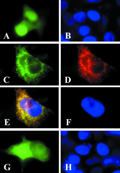
Figure 3.
The ACC2 N terminus directs the GFP to the mitochondria. Neonatal rat cardiomyocytes were plated at a density of 2 × 10⁵ on glass coverslips as described in the legend to Fig. 2. The cells were transfected with 1 μg/ml pEGFP-N1 (A and B), pEGFP-N1-ACC2-N (C and D), or pEGFP-N1-ACC1-N (G and H) by using the transfection reagent Lipofectamine according to the manufacturer's protocol. After 48 h of transfection, the cells were examined by fluorescence microscopy to verify expression of the GFP fusion protein (green). Cells that were transfected with pEGFP-N1-ACC2-N were immunostained with ACC2 antibodies and visualized by reacting them with the goat anti-rabbit IgG-TXRD conjugate (D). (E) A composite image of C and D; the overlapping regions in E are yellow. The cells were counterstained with DAPI (blue).

